
Secondary haemostasis and laboratory tests
Read the draft explanation
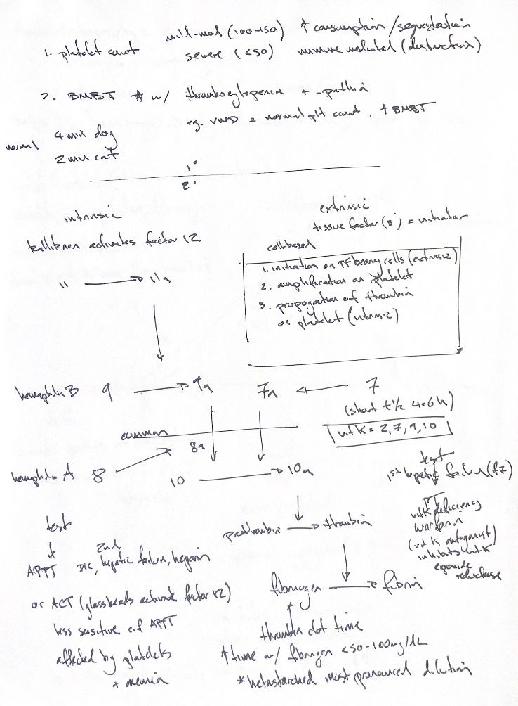
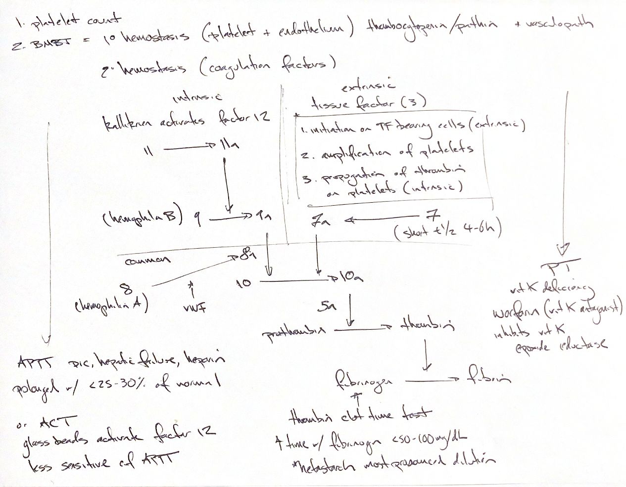
Platelet count checks number. Buccal mucosal bleeding time (BMBT) screens platelets, vWF and the vessel wall. Von Willebrand disease (vWD) may have a normal count but prolonged BMBT. Device and method matter. My dog 4-minute/cat 2-minute cues differ from this edition’s dog <3 minutes/sedated cat 34–105 seconds.
The cascade helps read plasma tests. aPTT covers intrinsic and common pathways; PT covers extrinsic and common. VIIIa works with IXa before X, despite its low position in my sketch. VIII deficiency causes haemophilia A; IX deficiency causes B. vWF carries and protects VIII.
In living blood, Tissue-factor-bearing cells make a little thrombin. It activates platelets and cofactors. Complexes on activated platelets then make a thrombin burst and fibrin. Initiation, amplification and propagation overlap.
Vitamin K supports II, VII, IX and X. Warfarin blocks vitamin K epoxide reductase. PT often prolongs first; the book gives VII a 4–6-hour half-life. Disseminated intravascular coagulation (DIC), hepatic failure and unfractionated heparin can prolong aPTT. The 25–30% cue is NORMAL REMAINING in an isolated factor defect, with reagent limits.
ACT uses whole blood and contact activators, including glass, to trigger XII. It is less sensitive than aPTT; platelets and anaemia affect it. XII deficiency can prolong tests without a bleeding tendency.
Thrombin clot time adds thrombin to test fibrinogen-to-fibrin conversion. Low/abnormal fibrinogen and inhibitors prolong it. The book’s <50–100 mg/dL describes severe depletion before routine times often prolong, not a universal TCT cutoff. One sketch appears to say mg/µL; the paired sketch and book use mg/dL. The original/book “hetastarch: most pronounced dilution” cue is context-dependent; effects vary with formulation and setting.
Read tests with the patient. They do not reliably predict surgical bleeding.
References
Johnston/Tobias 2nd ed., Ch.7; Alatzas 2014; Cornell eClinpath.