Start here: what is actually wrong
Cruciate disease affects the ligament, the joint, and sometimes the meniscus.
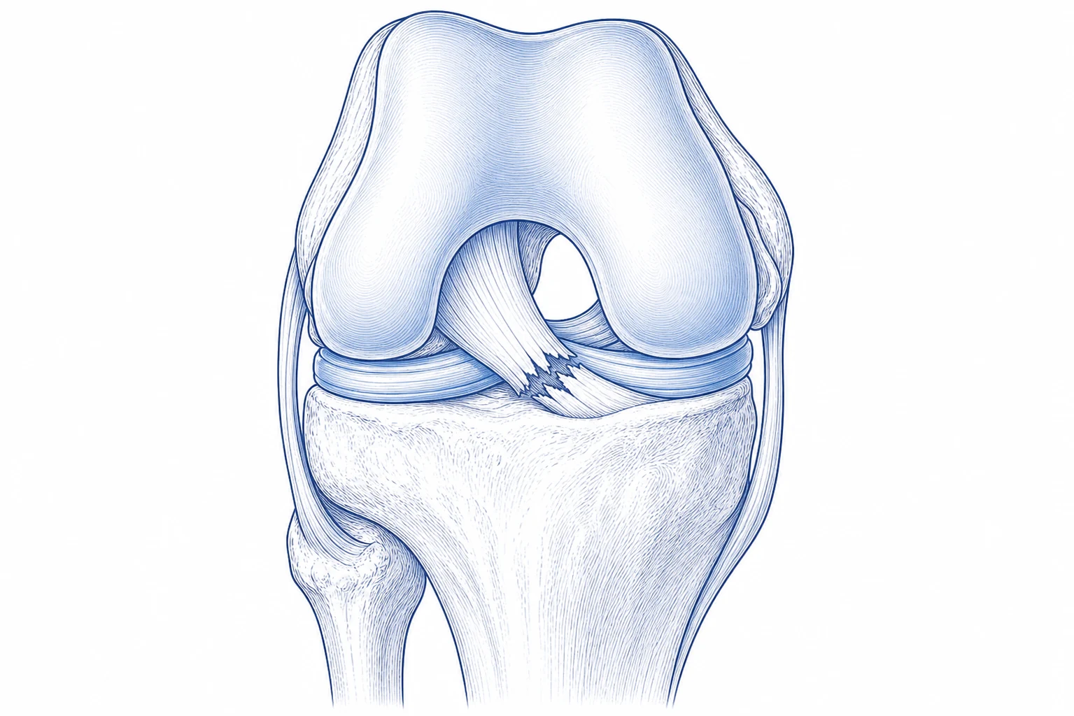
What cruciate disease meansCruciate disease is a common reason dogs develop knee pain and back-leg lameness.
Cruciate disease is one of the most common reasons dogs develop knee pain and back-leg lameness.
The cruciate ligament is one of the main stabilizers inside the knee. In many dogs, it does not simply snap during one accident. It often weakens gradually, a bit like a rope that is fraying. At some point it may partially tear or completely tear.
Once the ligament is no longer doing its job, the knee can move abnormally when your dog stands or walks. That abnormal motion creates pain and inflammation inside the joint, and over time it contributes to osteoarthritis.
What the meniscus isThe meniscus is a shock absorber that can be injured along with cruciate disease.
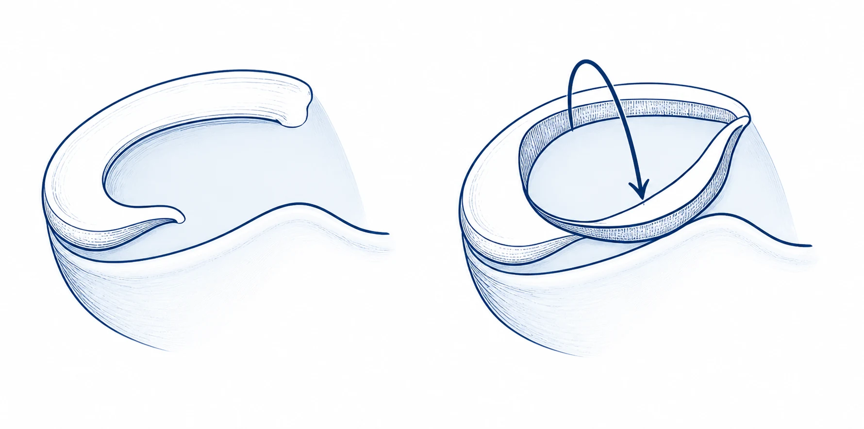
The meniscus is a C-shaped cushion, a bit like a shock absorber, inside the knee. It sits between the thigh bone and shin bone and helps distribute load when your dog stands, walks, and turns.
When the cruciate ligament is damaged, the knee can move abnormally. That abnormal motion can pinch or tear the meniscus. A meniscal tear can add pain, catching, clicking, or a more persistent limp.
A click can make us more suspicious of a meniscal tear, but many dogs with meniscal injury do not have an obvious click. If TPLO surgery is performed, the meniscus is checked during surgery and treated if needed.
How the consult confirms the problem
Diagnosis is usually based on the whole picture, not one single test.
What we examineThe exam looks at the whole knee, not just one sign.
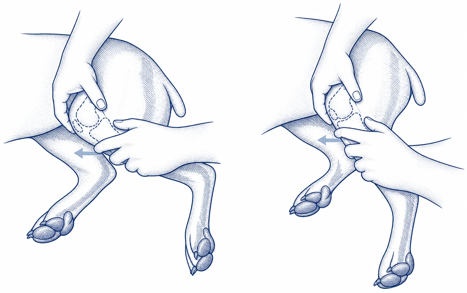
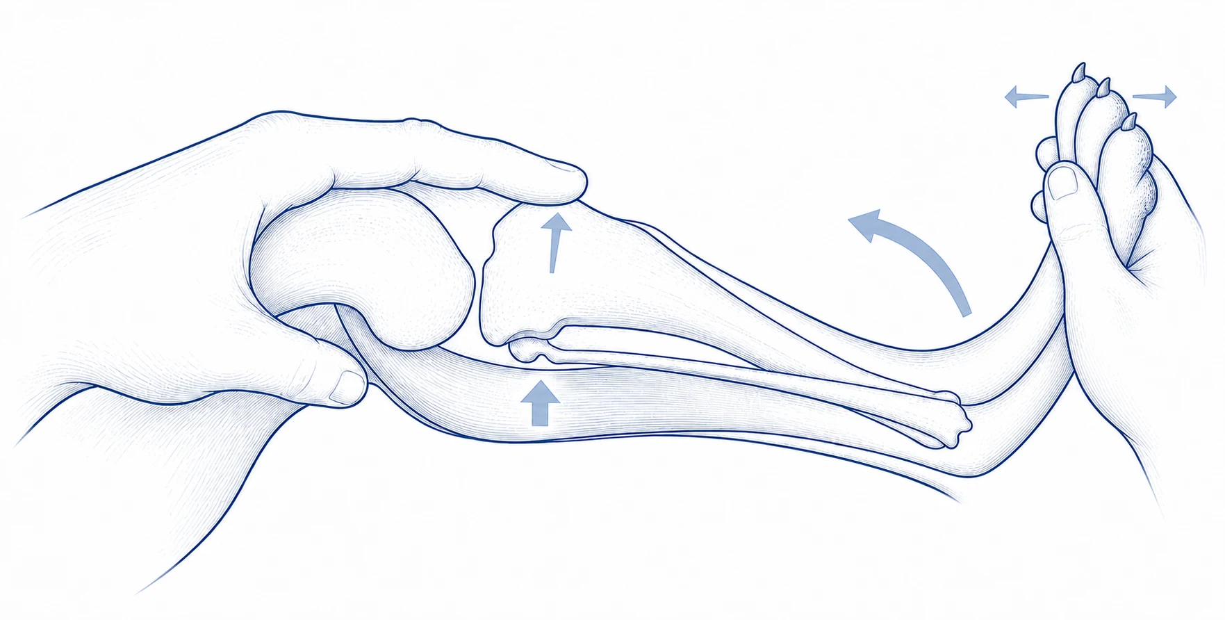
During the consult, I assess the whole picture: how your dog uses the leg, where the knee is painful, how the joint moves, and whether the knee shows instability.
- How your dog stands, walks, sits, turns, and uses the sore leg.
- Pain, swelling, thickening, range of motion, and comfort through the knee.
- Meniscal clues, such as clicking, catching, joint discomfort, or a limp that does not fit the rest of the exam.
- Standard cruciate instability checks, such as the ones shown here.
- Rotational or pivot instability, assessed with the tibial pivot compression test.
- The kneecap, hips, opposite knee, alignment, and body condition, because these can affect diagnosis, treatment planning, and recovery.
- Sedated examination may help when the physical exam is limited or unclear.
What imaging addsThe exam diagnoses instability; calibrated radiographs help rule out other problems and plan TPLO.
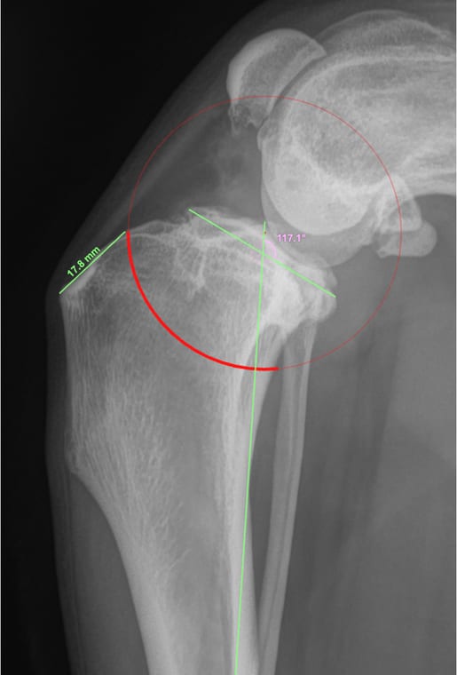
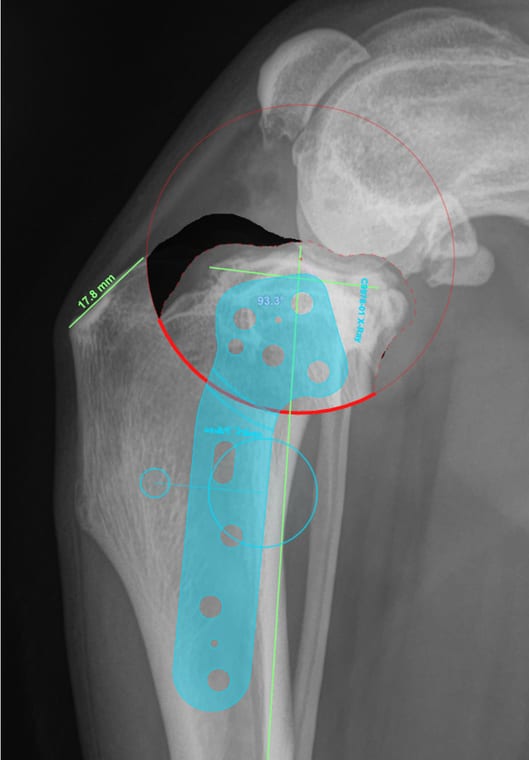
Radiographs help look for other causes of pain and show bone and joint changes around the knee.
The diagnosis of knee instability is primarily made on the orthopedic exam. Radiographs are still important, but they answer different questions: they help look for other causes of pain and provide the measurements needed for surgical planning.
- Radiographs can show arthritis, joint swelling, bone shape, and other changes around the knee, and help rule out other bone or joint problems, including bone disease or neoplasia, that could change the plan.
- For TPLO planning, the X-rays need a calibration marker, often a calibration ball, so measurements are accurate.
- Calibrated planning radiographs help choose plate size, screw lengths, saw blade size, cut placement, and how much the bone segment should be rotated.
- Radiographs do not directly show the cruciate ligament or every meniscal injury.
- Some details, such as meniscal injury or cartilage changes, may only be assessed more directly during surgery or joint inspection when available.
Does my dog still need TPLO?
A better day, a partial tear, or an older age does not answer this question by itself. The recommendation depends on pain, knee stability, the examination, imaging, overall health, and what your dog needs to do comfortably.
My dog is walking better. Does that mean surgery is no longer needed?Improvement in the limp does not necessarily mean that the ligament or knee stability has returned to normal.
Cruciate-disease lameness can vary from day to day. Rest and medication may improve comfort, and scar tissue may reduce some movement, but a dog walking better does not by itself show that the ligament has healed or that the knee is stable.
The decision should be based on the orthopedic examination, the amount and direction of instability, pain, imaging findings, activity needs, and the likely consequences of leaving the knee unstable—not on one good day or a short improvement in the limp.
Can a partial cruciate tear heal without surgery?Partial tears can be harder to diagnose and commonly progress, although treatment is still individualized.
A partial tear does not always produce obvious looseness during an awake examination, and the limp may temporarily improve. However, partial cruciate tearing commonly progresses over time.
Non-surgical care may be considered for selected dogs and can include activity modification, weight management, pain medication, and rehabilitation. It may improve comfort or delay surgery, but improvement does not prove that normal ligament strength or knee stability has returned. The examination and the dog's size, age, activity, anatomy, and degree of instability help determine whether continued non-surgical care or surgery is more appropriate.
Is my dog too old, too small, overweight, or not active enough for TPLO?Age or size alone does not decide the recommendation.
Cruciate disease affects dogs of many ages and sizes. I consider overall health and anesthetic risk, body weight and condition, knee instability, arthritis, other orthopedic problems, daily activity, home support, and the owner's goals.
An older or small dog may still be a TPLO candidate, while another dog may be better suited to a different operation or non-surgical management. Excess body weight increases the load on an affected knee, so a realistic weight plan is often part of treatment and recovery rather than a reason to dismiss the problem.
Why TPLO changes the knee mechanics
When the consult findings support cranial cruciate ligament disease as the source of painful knee instability, TPLO is usually the surgery discussed.
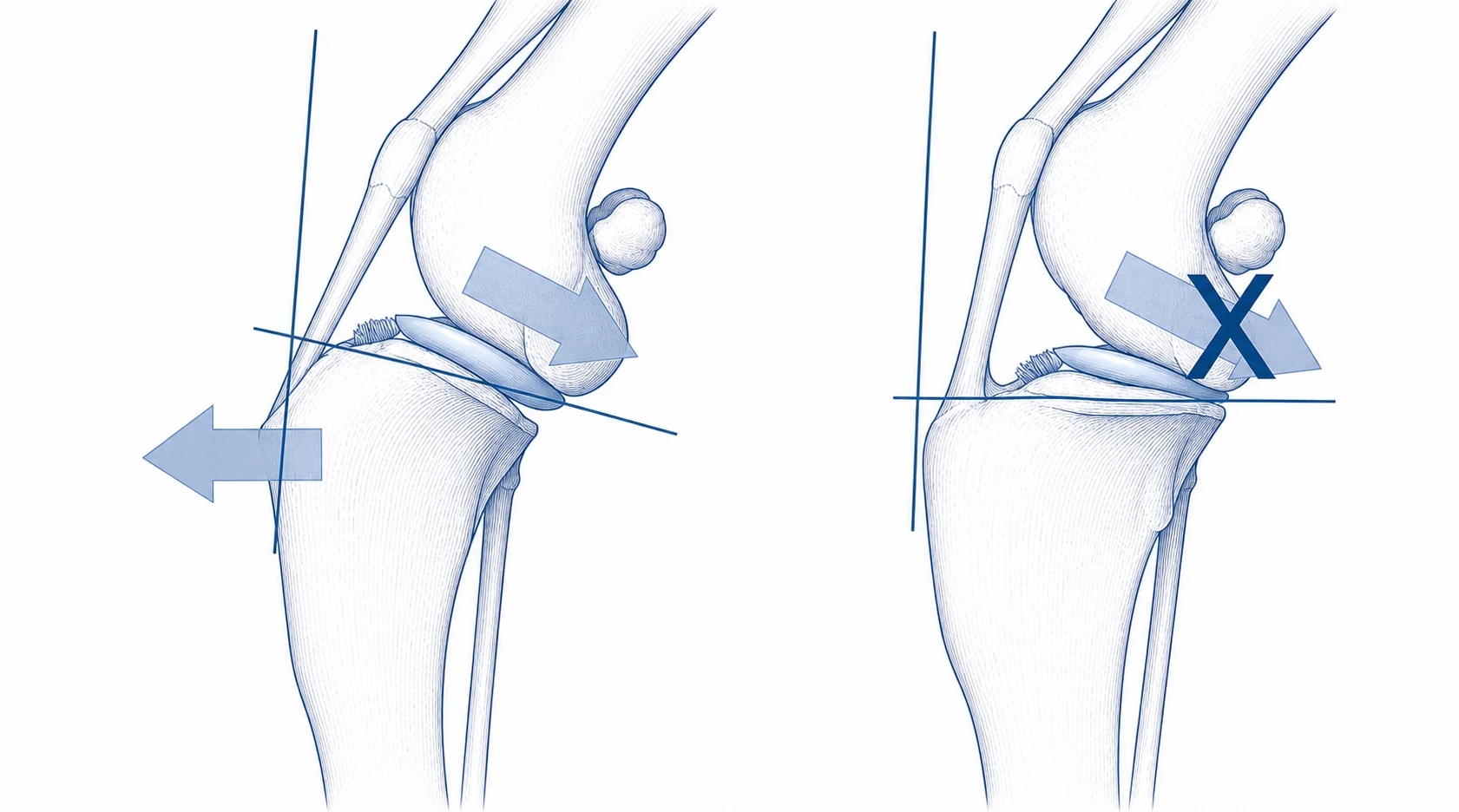
What the procedure changesThe procedure changes how the knee bears weight rather than replacing the ligament.
The cruciate ligament normally helps control abnormal sliding or shearing motion in the knee. When it is damaged, the knee becomes painful and unstable, especially when your dog bears weight.
TPLO stands for tibial plateau leveling osteotomy. Instead of replacing the torn cruciate ligament, TPLO changes the slope of the top of the tibia with a controlled curved bone cut. In owner terms, I often describe it as a controlled, carefully planned bone cut, almost like a controlled fracture. The bone segment is rotated into a new planned position and held with a plate and screws while it heals. This changes how the knee bears weight so it can be functionally more stable.
Muscle-sparing surgical techniqueThe surgical approach preserves key muscle attachments during TPLO.
Every TPLO is performed with a muscle-sparing, or pes-sparing, approach. The pes anserinus is where several hamstring and adductor muscle-tendon attachments meet on the inside of the upper shin bone, close to where TPLO is performed.
In a traditional TPLO exposure, this attachment may be lifted or cut from the tibia for access and then sutured back over the plate. That repaired layer must heal and can separate. My approach uses only a small opening through the fascia, preserves the muscle attachment to the bone, and plans the plate position around the preserved tissues from the beginning rather than treating them as an afterthought at closure.
Surgical photo comparisonIntra-operative views before closure; tap to compare exposure.
These before-closure images compare a traditional exposure with the muscle-sparing approach, showing the difference in how much of the muscle attachment is released to access the upper tibia.
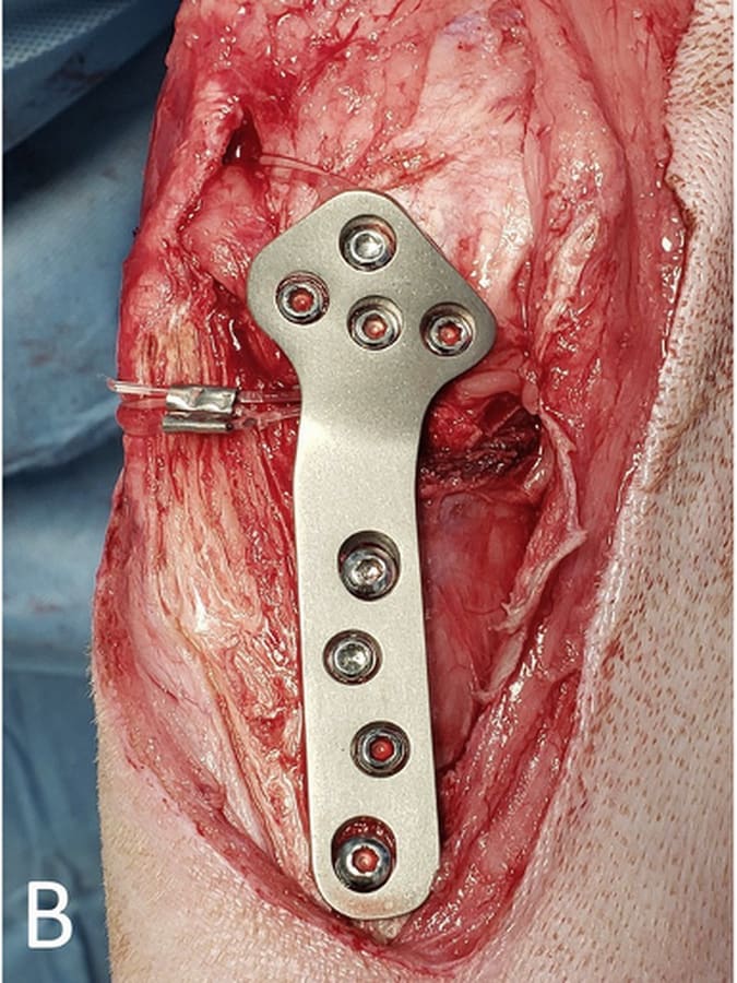
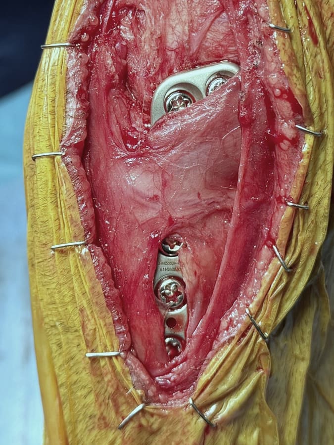
This approach is extrapolated from human high tibial osteotomy literature, where preserving the pes anserinus has been associated with maintained alignment correction, earlier bone healing, and better early function. The same principle guides the technique here: accurate TPLO surgery with deliberate protection of the muscle attachments around the bone.
What the procedure does not doThe procedure does not replace the torn ligament or remove all arthritis.
TPLO does not replace the torn ligament, and it does not remove all arthritis. It is a bone surgery: a controlled cut is made in the top of the shin bone, then the bone is stabilized while it heals.
What happens to the meniscus?TPLO helps the knee load the meniscus
in a more normal pattern.
The important point is that TPLO can change how pressure is carried through the meniscus. In studies that measure pressure inside the knee, the pattern after TPLO can look more like a knee before the cruciate ligament was torn than a knee with a torn ligament.
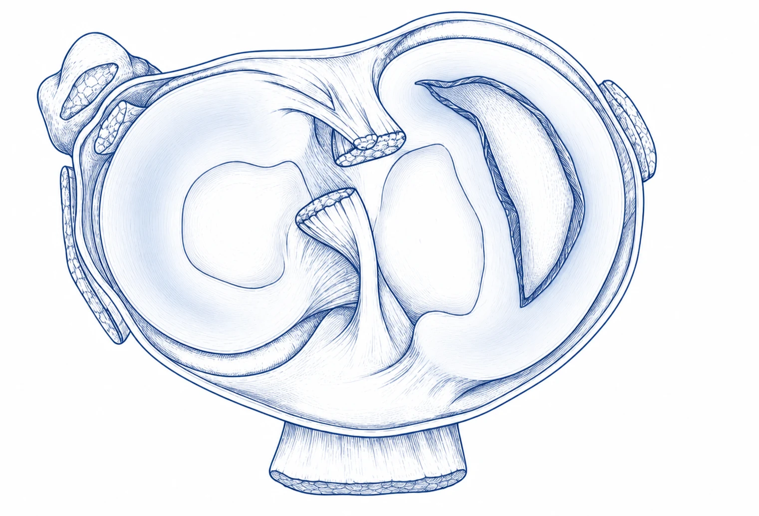
Pressure-map exampleHow knee loading changes before and after TPLO.
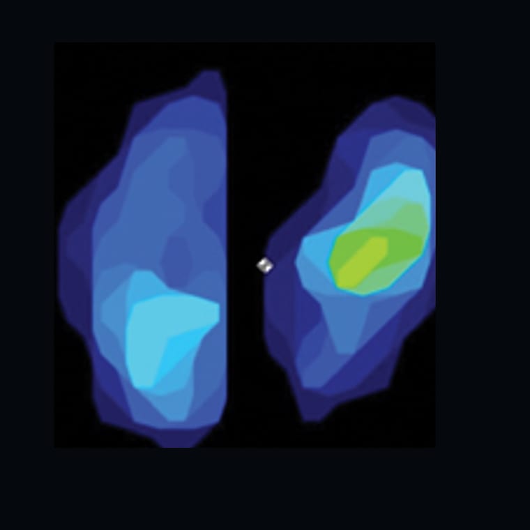
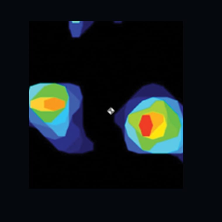
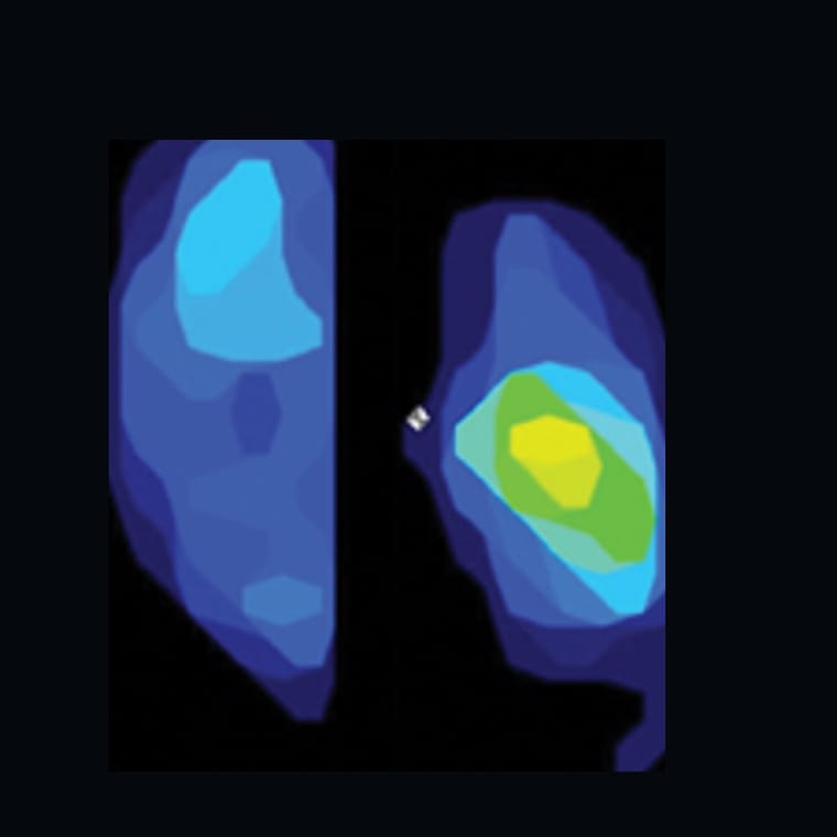
Torn ligament
TPLO changes the way weight passes through the knee. This can help reduce the abnormal pinching forces on the meniscus. That matters because meniscus that remains stable can still help cushion and protect the joint.
If there is a displaced bucket-handle tear that can catch in the joint and is considered a likely source of pain, the torn portion is removed. Otherwise, the meniscus is preserved.
How each TPLO is planned and supported
Dr. Chris Wood, BSc, DVM (Dist), MS, DACVS-SA, is an ACVS board-certified small animal veterinary surgeon in Ottawa with a professional interest in hip and stifle disorders, including TPLO.
For broader hip, stifle, fracture, and orthopedic referral information, review veterinary orthopedic surgery in Ottawa.
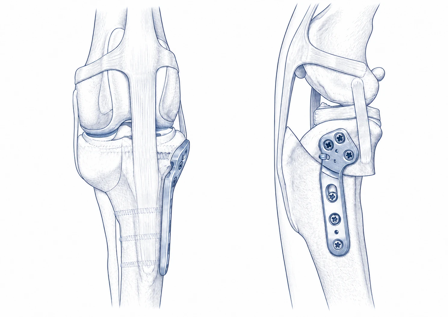
Dr Wood plans each TPLO from calibrated radiographs using VPOP, a virtual orthopedic planning program. He checks and refines the measurements and surgical plan for each dog before surgery.
A medial mini-arthrotomy is used to inspect the meniscus during every TPLO. The meniscus is preserved unless a displaced bucket-handle tear is considered a likely source of pain; when present, only the torn portion is removed.
Dr Wood uses a pes-preserving approach, leaving the pes anserinus attachment intact rather than releasing it from the tibia.
A board-certified veterinary anesthesiologist is involved in every TPLO. Dogs recover in a specialty and emergency hospital, with overnight monitoring available when needed.
Hospital stay, same-day discharge, and bilateral surgery
Hospitalization is planned around anesthetic recovery, comfort, mobility, and individual medical needs. For cost-conscious owners, same-day discharge—going home on the day of surgery—is available when the dog is medically comfortable and safe to leave the hospital. Dogs who need continued care stay overnight.
Single-session bilateral TPLO is available for selected dogs with disease affecting both knees. The current bilateral package includes a two-night hospital stay because the early care and mobility demands are greater.
There is no single plan that is best for every dog with two affected knees. One session means one anesthetic and one combined restriction period, but the first days require more help with standing, toileting, and movement. Staged surgery allows one leg to recover before the second operation, but it involves two anesthetics and two recovery periods. The decision considers the severity in each knee, body size and condition, other health or orthopedic problems, mobility, home layout, and the help available during recovery.
When appropriate, the surgery team can help a referring veterinarian obtain suitable calibrated planning radiographs locally. This may reduce duplicated imaging, travel, or cost, although images may need to be repeated if they are not adequate for surgical planning.
Recovery planning for your home and routine
Before surgery, it helps to picture the home routine recovery will require. The early goal is a quiet, grippy, predictable setup: incision protection, leash-only toileting, medication monitoring, and preventing slipping, jumping, running, rough play, or free yard access.
First 10 to 14 daysIncision checks, cone or incision protection, toileting-only leash walks, medications, and preventing jumping or rough activity.
Early recovery focuses on incision protection, cone or recovery-collar use, toileting-only leash walks, pain control, medication monitoring, and preventing jumping, slipping, running, rough play, or off-leash activity. Some soreness, swelling, bruising, tiredness, or mild lameness can occur early, but the overall trend should be toward comfort and controlled limb use.
Early leash-walk weeksWalking targets, simple exercises, and stair access are staged rather than rushed.
The rehab plan adds walking targets and simple exercises by stage. Stairs are treated as controlled household access, not exercise. Running, jumping, trotting, rough play, sport, and off-leash activity wait until healing has been assessed and the next stage has been cleared.
Healing check and recheck X-raysRecheck X-rays are planned at 8–10 weeks to assess bone healing before activity advances.
Recheck X-rays are planned at 8–10 weeks to assess bone healing and implant position before activity is advanced. The exact date within that window can change depending on the dog, comfort, progress, and surgeon review.
The TPLO recovery guide includes the detailed Week 8 recheck plan and after-clearance activity progression.
Gradual return after clearanceActivity increases after clinical and radiographic healing, not just because the limp looks better.
After radiographic and clinical healing, activity is gradually increased. Running, jumping, stair exercise, rough play, and sport should wait until cleared.
TPLO estimate range
Final written estimates are prepared after the consult, examination, imaging review, and surgical plan.
$5,800-8,000 pre-tax
- TPLO with internal brace augmentation.
- TPLO with patellar luxation correction.
$7,800-10,500 pre-tax
- Bilateral single-session TPLO.
Estimate after consult
- Angular limb deformity correction.
- Additional joint reconstruction or unexpected aftercare needs.
The consult can focus on your dog's exam, imaging, home routine, recovery constraints, and goals. The final plan and written estimate are made after that assessment.