
Primary haemostasis and endothelial platelet inhibition
Read the draft explanation
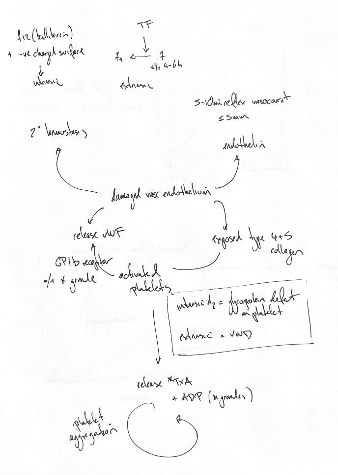
An injured vessel exposes collagen and von Willebrand factor (vWF). Platelets use GPIb to bind vWF and attach at the injury. Other receptors also help them bind collagen. Activation then brings more platelets into the growing plug.
Activated platelets release ADP from dense granules. They make thromboxane A2 from arachidonic acid. These signals recruit more platelets. Activated integrin αIIbβ3 binds fibrinogen, which bridges neighbouring platelets. My original ADP “alpha-granule” annotation is corrected here: ADP is stored in dense granules. vWF is an alpha-granule protein, with species differences in its platelet storage.
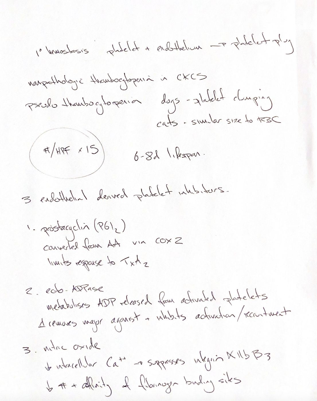
Healthy endothelium limits this response in three ways. Prostacyclin (PGI2) restrains activation. Surface ecto-ADPase breaks down the released ADP signal. Nitric oxide lowers platelet calcium signalling and limits activation of the fibrinogen receptor. My notes and the book use a COX-2 cue for PGI2. Isoform contributions depend on the setting; this is not an exclusive rule for every species.
A low automated platelet count needs a smear check. Clumps and large platelets can cause an undercount. Some Cavalier King Charles Spaniels have benign inherited macrothrombocytopenia, but breed alone does not exclude disease. The smear shortcut uses the average count across 10 oil-immersion fields ×15,000/µL. Clumping makes that estimate unreliable.
Primary haemostasis builds the platelet plug while coagulation develops alongside it. The source labels “intrinsic” and “extrinsic” for platelet-receptor defects versus vWF deficiency describe the site of a primary defect. They are separate from the coagulation-pathway names shown on the map.
References
Johnston/Tobias, 2nd ed.; Cornell eClinPath; Kirkby and Barbieri 2012 (selected primary abstracts).