
Lumps and tumours
A lump does not always mean cancer. Learn how the surgical team assesses lumps and plans treatment, or go straight to the guide for your pet’s diagnosis or the location of the lump.
Choose a condition to see what to send with the referral.
Send clinical notes, pathology and blood results, plus original images and reports. Further tests are listed under “Coordinate with the surgical team”.
From finding a lump to a treatment plan
Overview
No diagnosis yet? You can ask for an assessment without choosing a tumour type. A lump is not automatically cancer, and its appearance alone cannot identify it.
The first assessment starts with an examination and any samples already taken. The next questions are whether the lump needs treatment, which tests could help, and what an operation would involve. Some lumps can be monitored; others need removal or a different treatment plan.
Have a diagnosis or know the location? Choose your pet’s guide →
Tests and planning
What needs checking?
- What is the lump? A fine-needle aspirate collects cells through a small needle. Some lumps need a biopsy—a piece of tissue examined in the laboratory. The biopsy site is planned so it can be included in later surgery.
- How far does it extend, and has it spread? Selected X-rays, ultrasound or CT scans can show how the lump involves nearby tissues and check for visible spread to other parts of the body. Assessing the extent of cancer is called staging.
- Does a lymph node need checking? These small structures filter fluid from tissues. For selected tumours, mapping follows drainage to find which node to sample. The laboratory examines cells or tissue to find out whether cancer is present.
How could spread change the operation?
If cancer has spread elsewhere, a wider removal around the original lump may add a larger wound and complication risk without enough extra benefit. The aim of surgery may change from lasting tumour control to relief of pain, bleeding or another problem. Discuss whether that benefit justifies the operation and recovery with the surgical team.
Spread to a lymph node does not automatically rule out surgery. The tumour type, extent of disease and your pet’s health all matter. The team recommends tests when the results could change care. Normal scans cannot rule out microscopic disease.
Finding the right lymph node to test
Mapping is not needed for every lump. It follows drainage to find which node to check; this is not always the nearest one.
Read how sentinel lymph node mapping works →
Bring existing sample reports, scan reports and details of previous surgery. Tell the surgical team if the lump has already been removed.
Surgery
Why can the wound be larger than the lump?
The plan may include tissue around and beneath the lump. How much can be removed depends on the suspected tumour, its location and nearby structures. Surgical planning also covers wound closure and protecting movement.
This is the plan before surgery. The laboratory checks the cut edges afterward, as explained under Results and next steps.
Closer or wider removal: what the terms mean
For some tumours, closer removal may preserve movement or make closure safer, while increasing the chance that tumour cells remain. Discuss that trade-off with the surgical team before surgery.
How is the wound closed?
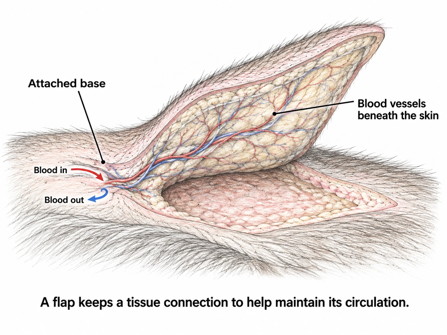
Some wounds close with nearby loose skin. Others need a skin flap, which moves neighbouring skin while keeping a tissue connection. Discuss the likely wound and its care with the surgical team before surgery.
If the lump was removed before referral, the earlier operation and tissue report help determine whether tissue around the scar needs further surgery.
Results and next steps
The laboratory identifies the removed tissue and may report a grade: features seen under the microscope that help judge a tumour’s likely behaviour. Grade is different from stage, which describes the disease’s extent, including evidence of spread.
The margins are the cut edges of the removed tissue. If tumour cells reach an edge, some may remain in the wound. A clear edge is useful information, but cannot guarantee that a tumour will not return.
The surgical team reviews the tissue report, any lymph-node results and scans together. Depending on the findings, the next step may be monitoring, further surgery, or discussion with a cancer care team about radiation or medicines. Not every pet needs further treatment.
Recovery
Follow your pet’s written discharge plan for medicines, activity, wound protection and rechecks. Prevent licking or chewing and check the wound each day. Any bandage or drain has its own care instructions.
Contact the team for wound opening, bleeding, discharge, increasing pain, or spreading redness or swelling. A new swelling should be assessed rather than assumed to be fluid.
Use the contact details on your discharge sheet. If you do not have them, contact your treating veterinary team. For an urgent or worsening concern, call rather than waiting for an email reply.
Read the wound-care guide → Your pet’s own instructions take priority.
Wound changes to report
The surgical team reviews the tissue results and wound healing before deciding what comes next. Keep the recommended rechecks, and ask your vet to assess a new lump or a change at the scar.
Find your pet’s guide
Choose a diagnosis or the location of the lump. You can read each guide on its own.
Sentinel lymph node mapping: finding the right node to test →
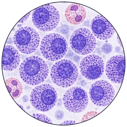 01Mast cell tumourTests, surgery and follow-up
This guide is about mast cell tumours in or under a dog’s skin. Mast cell disease in cats or in internal organs needs a different assessment.
Overview
What is a mast cell tumour?
Mast cells are part of the body’s defences, called the immune system. They contain chemicals that can cause swelling and irritation when released. A mast cell tumour forms when these cells grow out of control. You may see the name shortened to MCT on your dog’s test results.
These tumours can look like many other skin lumps. Some grow slowly and can be controlled with surgery alone. Others grow into nearby tissue or spread elsewhere in the body. A lump’s size or appearance cannot show how it will behave.
What needs checking?
A small needle sample can often identify the lump. The sample helps guide further tests and treatment, including how to close the wound if the lump is removed.
Testing a piece of the lump, or the whole lump after removal, provides a tissue report with more information about its likely behaviour. Some treatment decisions need to wait for the tissue results.
Signs and symptoms
What you might notice
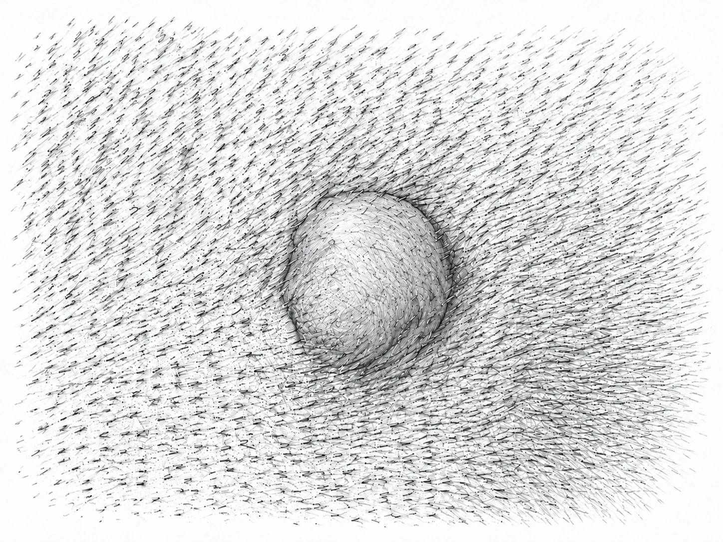
A mast cell tumour may be a small lump in the skin or a deeper swelling beneath it. It may have normal skin over it, or look red, bruised or sore. Some develop an open or bleeding surface. A dog with a small tumour may otherwise seem well.
The lump can swell and then shrink. This can happen when the tumour cells release chemicals that irritate the nearby tissue. Shrinking does not mean the tumour has gone away. Avoid squeezing or repeatedly rubbing it.
Changes to tell your vet about
Tell your vet when you first noticed the lump, how it has changed, and whether your dog has had a lump removed from that spot before. Mention any other lumps, loss of appetite or vomiting. Photos and measurements can help show changes over time.
Call your vet promptly for repeated vomiting or black, tar-like stool. If your dog collapses or becomes very weak, seek emergency care immediately. Mast cell tumours can sometimes cause stomach ulcers—sores in the stomach lining—or other serious effects. These signs can also have other causes and need veterinary assessment.
Diagnosis
Start with a needle sample
A fine-needle aspirate, or FNA, uses a small needle to collect cells from the lump. The cells are examined under a microscope. This can often identify a mast cell tumour without removing the whole lump.
The sample may show features that raise concern, but it does not give the final tissue grade. If the sample is unclear, or knowing more would change the operation, the surgical team may recommend another sample or a biopsy. A biopsy removes a piece of tissue for testing.
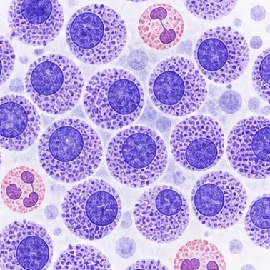
Grade and stage answer different questions
- Grade: how the tumour cells and tissue look under a microscope. These findings help show how the tumour is likely to behave. It comes from tissue examined after a biopsy or removal.
- Stage: the extent of the disease, including the tumour’s size and whether it has spread to lymph nodes or other organs. Examination, lymph-node samples and selected scans help assess this.
For tumours within a dog’s skin, the report may use low-grade or high-grade, or an older system with grades I, II and III. These systems do not match exactly. Tumours arising beneath the skin are assessed differently. The surgical team will explain what your dog’s report means.
Why check a lymph node?
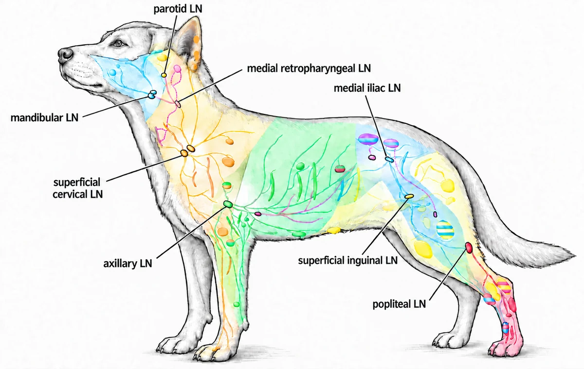
Lymph nodes are small structures that filter fluid draining from tissues. They can be an early site of tumour spread. A node can contain tumour cells even when it feels normal in size.
Sentinel lymph-node mapping finds which node or nodes receive drainage from the tumour area. They are not always the nearest ones. Mapping finds the node to test; it does not show whether cancer is present. That requires examining cells or tissue from the node.
How sentinel lymph node mapping works →
General lymphatic anatomy in a dog
Does every dog need the same scans?
No. The tests depend on the lump, your dog’s examination and health, and whether the results would change treatment. The surgical team may recommend an ultrasound and samples from the liver or spleen when there is more concern about spread. Chest imaging or a scan of the tumour area may be useful for a specific concern. How samples, scans and lymph-node tests help the plan →
Having several skin lumps does not by itself prove that one tumour has spread. Each lump still needs assessment. If a lump has already been removed, send the tissue report and details of the surgery to the surgical team.
Treatment
Planning the operation
Surgery is the main treatment for many mast cell tumours in the skin. Dr. Wood plans to remove the tumour with tissue around and beneath it, while protecting movement and leaving a wound that can heal.
The wound may be larger than the lump you can see because the removal includes tissue around and beneath it. On a leg or the face there may be less spare skin; some wounds need nearby skin moved to close the gap. Discuss the planned removal and closure with the surgical team before surgery. Why removal can leave a larger wound and how it can be closed →
How nearby skin can help close a gap
Dr. Wood’s usual plan includes sentinel lymph-node mapping and removal of the node for testing, along with the mass. If medicine is needed to control the effects of chemicals released by the tumour, the surgical team will give you specific instructions.
What the tissue report shows
The laboratory examines the tumour and its cut edges, called the margins. Tumour cells at an edge suggest some may remain in the wound. A clear edge is useful, but cannot guarantee the tumour will not return. What the tissue report adds to the plan →
The surgical team considers the margins, grade and any evidence of spread together. The next step may be monitoring, another operation to remove tissue around the scar, or discussion with an oncology team about radiation or medicines. Radiation treats a chosen area; some cancer medicines treat disease elsewhere in the body. Not every dog needs these treatments.
Before booking surgery, ask the surgical team about the expected hospital stay, help your pet may need at home and planned rechecks.
Aftercare and outcome
Looking after the wound
Home care depends on where the tumour was removed, how the wound was closed, and whether a lymph node was removed too. Follow your dog’s written discharge instructions for activity, medicines, wound protection and rechecks.
Keep your dog from licking or chewing the wound using the protection recommended by the surgery team. Check the wound each day. Contact the hospital if it opens, bleeds, develops discharge, becomes more painful, or has spreading redness or swelling.
Use the contact details on your discharge sheet. If you do not have them, contact your treating veterinary team. For an urgent or worsening concern, call rather than waiting for an email reply.
If your dog becomes suddenly unwell, has repeated vomiting or black stool, call rather than waiting for an email reply. The shared recovery guide explains the home-care plan and links to practical wound-care advice. Your dog’s own instructions take priority.
Reviewing the results
The tissue and lymph-node results help determine whether further treatment is needed. The team also checks wound healing before starting more treatment.
Low-grade tumours have tissue features linked to less aggressive behaviour. Many low-grade skin mast cell tumours do well after suitable surgery. Higher-grade tumours, spread to a lymph node, or tumour left behind may change the plan. The needle sample or the lump’s appearance alone cannot give a reliable outlook.
Checking for new lumps
Keep checking the scar and the rest of your dog’s skin. Ask your vet to assess a new lump or a change at the scar. A new mast cell tumour elsewhere can be a separate tumour; it does not automatically mean the first one has spread.
For veterinary teams Mast cell tumour referral checklist
← Back to the owner explanation
Mast cell tumour: referral checklist
Before referral
- Mass FNA — include cytology/pathology report
- CBC / biochemistry
- Site, size (3 dimensions), depth/mobility, lymph-node findings and photos
- Growth, ulceration, recurrence/prior surgery, medications and systemic/GI signs
- Chest imaging: not routine for low-risk cutaneous MCT. Consider for systemic signs, thoracic concerns or facial/oral/perineal/preputial tumours if results would change management.
Send: blood results, any histopathology and original imaging/reports.
Coordinate with the surgical team
- Sentinel-node mapping / sampling as part of tumour surgery
- Abdominal US ± liver/spleen aspirates, if indicated
- Local imaging or biopsy, if indicated
Do not delay the tumour referral for mapping. Discuss feline or visceral MCT.
Prepare a referral →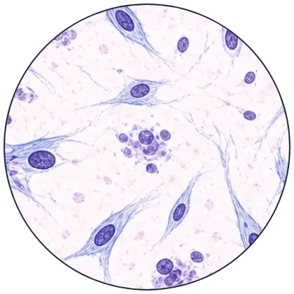 02Soft-tissue sarcomaTests and surgery for a soft-tissue cancer
This guide covers soft-tissue sarcomas in or under a dog’s skin. Tumours in internal organs, around major nerves, or at an injection site in a cat need a different assessment.
Overview
What is a soft-tissue sarcoma?
A soft-tissue sarcoma is a cancer that starts in tissues that support or connect other parts of the body. Several tumour types fall into this group. You may see the name shortened to STS on a test report.
These tumours can send tiny extensions into the tissue around the lump. Those cells are too small to see or feel during an examination. Removing only the visible lump may leave some behind, allowing it to grow back.
Has it grown nearby or spread elsewhere?
For many skin sarcomas, preventing regrowth at the original site is the main concern. Assessment also checks whether it may have spread elsewhere. The risk of spread depends on the tumour type and tissue test results.
Signs and symptoms
What you might notice
A sarcoma may feel like a lump under normal skin. Some grow slowly and cause no obvious pain. Others grow quickly, feel firmly attached, or affect how your dog walks. A larger tumour may develop a sore or bleeding surface. These changes cannot identify the tumour type on their own.
Tell your vet when you first noticed the lump, how it has changed, and whether anything was removed from that spot before. Bring photos or measurements if you have them. Arrange a prompt assessment for rapid growth, pain, an open surface or a change in movement.
Diagnosis
Why a needle sample may not give the answer
A fine-needle aspirate, or FNA, collects cells through a small needle. It can identify some other kinds of lump or suggest a sarcoma. Sarcomas may release few cells into the sample, so an unclear result does not rule one out.
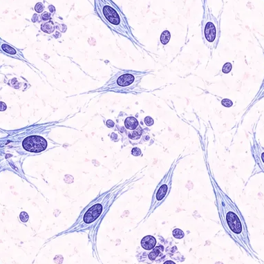
A biopsy takes a piece of tissue for laboratory testing. The surgical team may recommend one before removal if the result could change the operation or your decision about treatment. The biopsy site and any scar need planning so they can be included in the later removal. How samples and scans are selected →
What can the other tests show?
A tissue report gives the tumour’s type and grade. Grade describes features seen under the microscope that help predict its behaviour. It cannot be reliably assigned by feeling the lump or from a needle sample.
Chest X-rays or a CT scan check for visible spread to the lungs. CT uses X-rays to create detailed views inside the body. A scan of the lump itself may help plan surgery if it is deep, fixed, large or growing near important structures. No scan can rule out every microscopic tumour cell.
Treatment
Plan before removing the lump
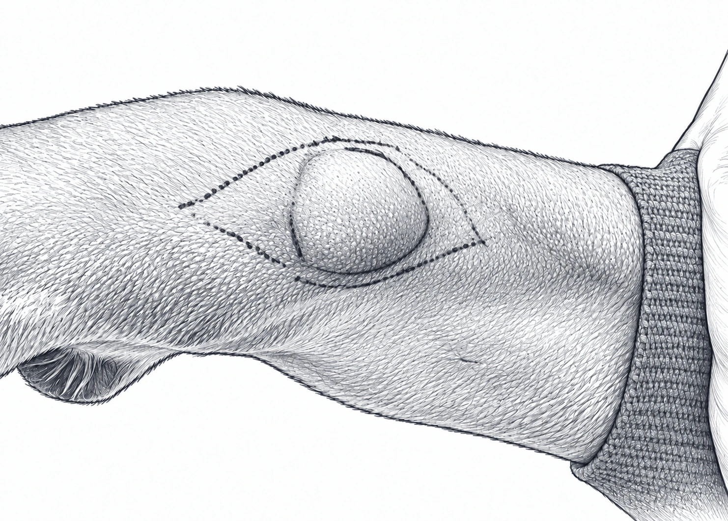
Surgery aims to remove the tumour with tissue around and beneath it, including the planned biopsy site or previous scar where needed. Tiny extensions may go beyond the part of the lump visible or felt on examination, so the wound may be much larger than the lump.
How will the wound be closed?
The tumour’s location affects how much tissue can be removed safely. The team plans wound closure before surgery. Some wounds need nearby skin moved over the gap. Read the shared removal and wound-closure explanation →
For some tumours, a smaller planned removal may preserve movement or make wound closure safer. It can also increase the chance that tumour cells remain and further treatment is needed. Discuss that trade-off with the surgical team before surgery.
Closer or wider removal: what the terms mean
One way a flap moves nearby skin
Why keeping a skin connection matters
What if tumour cells reach an edge?
The laboratory checks the edges of the removed tissue, called the margins. Tumour cells at an edge suggest some may remain in the wound. Depending on the grade, location and surgery already done, the surgical team may discuss another operation, radiation treatment to the area, or monitoring. Cancer medicines, called chemotherapy, are considered for selected tumours with greater risk of spread. They are not needed for every sarcoma.
Before booking surgery, ask the surgical team about the expected hospital stay, help your pet may need at home and planned rechecks.
Aftercare and outcome
Recovery and the tissue report
Follow the written discharge plan for activity, medicines and wound protection. Contact the hospital for wound opening, discharge, increasing pain or spreading swelling. The shared recovery guide covers the home-care plan and practical wound-care advice.
Use the contact details on your discharge sheet. If you do not have them, contact your treating veterinary team. For an urgent or worsening concern, call rather than waiting for an email reply.
The surgical team reviews the grade and margins with you before deciding what comes next. Many lower-grade tumours do well after suitable surgery. A higher grade, deeper growth, recurrence or tumour left behind may change the outlook and treatment plan. Clear margins are helpful, but cannot guarantee that a tumour will never return.
Check the scar for a new lump or thickening and keep the recommended rechecks. Further chest imaging depends on the tumour’s grade and risk of spread.
For veterinary teams Soft-tissue sarcoma referral checklist
Soft-tissue sarcoma: referral checklist
Before referral
- Mass FNA — include cytology/pathology report
- 3-view thoracic radiographs or thoracic CT — originals and report
- Site, size (3 dimensions), depth, mobility/fixation and photos
- Growth, pain, recurrence; previous excision and scar/biopsy-tract location
Send: any histopathology, operative notes and existing blood results.
Coordinate with the surgical team
- Core/incisional biopsy if diagnosis changes planning; avoid unplanned excision
- Local CT/MRI/US: >5 cm, deep/fixed, recurrent or anatomically constrained mass
- Abdominal imaging only for systemic signs, abdominal location or subtype-specific concerns
Discuss suspected feline injection-site sarcoma before biopsy or excision.
Prepare a referral →Planning notes and references
AAHA diagnostic and staging guidance
CT and biopsy planning
When contrast CT may be recommended
Contrast-enhanced CT is Dr Wood's usual additional local planning test when a mass is over 5 cm, deep, fixed, poorly defined, recurrent, close to a joint or an important nerve or blood vessel, or when wide margins in that location may require a flap, graft, or other closure plan.
CT shows the relationship between the mass and the tissues that may need to be removed or reconstructed. It does not provide a tumour grade.
When a preoperative biopsy may be recommended
A deliberately placed core or incisional biopsy may be useful for a distal-limb or other constrained mass when the result could change the margins, reconstruction, or whether to proceed with an operation that has substantial functional impact. The biopsy tract must be positioned so it can be removed with the definitive surgery.
Biopsy grade is not definitive. In a study of 68 dogs, the preoperative biopsy grade matched the complete-excision grade in 59% of soft-tissue sarcomas. It underestimated the final grade in 29% and overestimated it in 12%.
68-dog biopsy-grade study · spindle-cell sarcoma cytology study
 03Lipoma / fatty lumpWhen a fatty lump needs monitoring, imaging or removal
This guide is mainly about fatty lumps in dogs. A soft lump is not necessarily a lipoma; examination and sampling come first.
Overview
What is a lipoma?
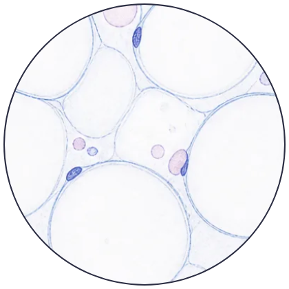
A lipoma is a benign tumour made of fat cells. Benign means it does not spread to other parts of the body like a cancer can. Many are simple lumps just under the skin. If testing and examination agree that a lump is a typical lipoma, it may only need monitoring.
Why does its location matter?
A fatty lump between muscles is called an intermuscular lipoma. It can grow quite large and interfere with movement even though it is benign.
An infiltrative lipoma grows into nearby tissues, such as muscle. It does not spread elsewhere in the body, but can be harder to remove and may grow back. A liposarcoma is a separate, cancerous tumour of fat tissue. Assessment and treatment follow the approach for sarcoma.
Signs and symptoms
What you might notice
A simple lipoma often feels soft and moves under the skin. The skin over it may look normal, and your dog may seem comfortable. Some dogs have several. Each new lump still needs its own assessment because different tumour types can feel alike.
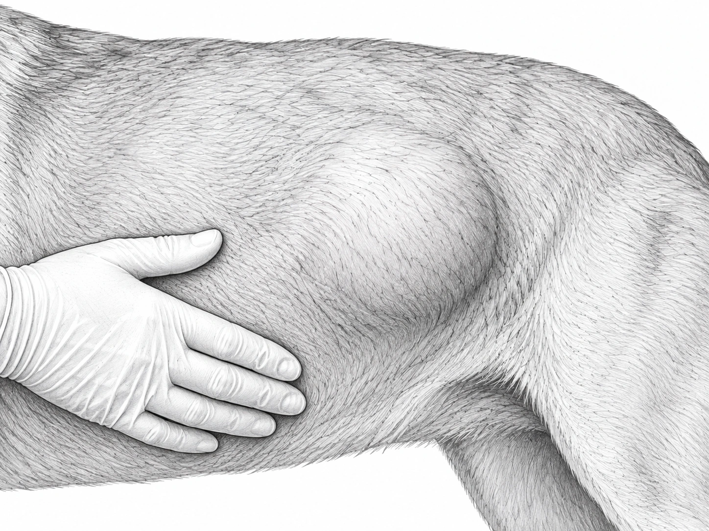
Tell your vet about rapid growth, pain, firmness, a lump that no longer moves freely, or a change in walking. A deep lump may cause a broad swelling rather than a small, easy-to-outline bump. These changes need assessment, but do not prove cancer.
Photos and measurements can help track change. Ask your vet to record which lump was sampled so a later change can be checked in the same place.
Diagnosis
Start with examination and a cell sample
A fine-needle aspirate, or FNA, uses a small needle to collect cells. Finding mature fat cells supports a lipoma diagnosis when the sample and examination fit together. Normal tissue also contains fat, so the sample needs to come from the lump being assessed.
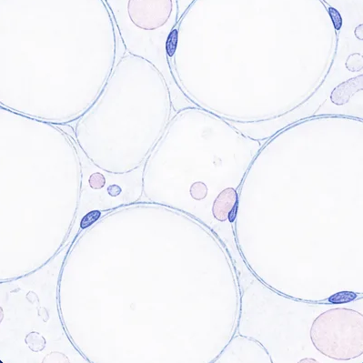
A needle sample does not show how far a fatty growth extends between or into muscles. If the lump is deep, fixed, painful, growing quickly or unlike a routine lipoma, a biopsy may be needed. A biopsy takes tissue for laboratory testing.
When is a scan useful?
A CT scan uses X-rays to build detailed views inside the body. CT or another scan may help show a deep lump’s extent and its relationship to muscles, nerves and blood vessels. The team recommends imaging when it could change the operation; the shared tests guide explains what different tests add. For a deep or unusual lump, the scan and any tissue results are considered together.
Routine chest or abdominal scans are not usually needed for a typical lipoma. If the findings suggest a sarcoma instead, the tests change.
Treatment
Monitor or remove?
A comfortable, typical lipoma may be monitored. Removal may make sense if it is growing, affecting movement, being rubbed or injured, or leaving uncertainty about the diagnosis. A growing lump in a tight space may become harder to remove later.
A simple lump under the skin and one deep among muscles can require very different operations. Before recommending removal, the surgical team assesses the planned removal and wound closure, nearby nerves and vessels, and how surgery may affect your dog’s movement.
Infiltrative lipomas need a separate plan
An infiltrative lipoma blends into nearby tissues, so removing every part may be difficult. Discuss with the surgical team whether surgery can remove it safely, whether some may remain, and the risk of regrowth. Radiation treatment to the area may be considered for selected tumours that cannot be removed or keep returning. It is not routine treatment for a simple lipoma.
Before booking surgery, ask the surgical team about the expected hospital stay, help your pet may need at home and planned rechecks.
Aftercare and outcome
What recovery involves
Removing a large fatty lump can leave a space under the skin where fluid collects. This is called a seroma. Some dogs need a drain to help fluid leave that space. Your discharge instructions cover the wound, any drain, medicines and activity.
Prevent licking and contact the hospital for increasing swelling, pain, discharge or wound opening. Do not assume a new swelling is just fluid. The shared recovery guide covers home care and changes to report.
Use the contact details on your discharge sheet. If you do not have them, contact your treating veterinary team. For an urgent or worsening concern, call rather than waiting for an email reply.
Can it grow back?
Simple and between-muscle lipomas usually do well after removal. Infiltrative lipomas have a greater risk of regrowth if tissue remains. The final tissue report and the operation’s findings guide follow-up.
Check the scar and have new lumps assessed. Another fatty lump elsewhere can be a separate lipoma; it does not mean the first one has spread.
For veterinary teams Lipoma referral checklist
Lipoma / fatty lump: referral checklist
Before referral
- Mass FNA — include cytology/pathology report
- Site, size (3 dimensions), depth, mobility/fixation and photos
- Growth, pain, altered movement/neurologic signs, prior surgery or recurrence
- No routine chest/abdominal staging for simple or infiltrative lipoma. Suspected liposarcoma/STS: thoracic staging before referral.
Send: any histopathology, operative notes, blood results and original imaging/reports.
Coordinate with the surgical team
- Local CT/MRI/US: deep, fixed, infiltrative, recurrent or function-limiting mass
- Biopsy for atypical mass or discordant examination/cytology
Planning notes and references
AAHA diagnostic and staging guidance
CT may be recommended for a deep or anatomically complex lipoma
Why CT and 3D planning may be useful
For a large, deep, intramuscular, recurrent, fixed, or anatomically complex lipoma, CT can map the muscle compartments, vessels, and the expected or visible course of important nerves. Dr Wood reviews the CT and performs his own 3D reconstruction for surgical planning.
CT can also suggest an infiltrative pattern. Within a CT comparison of 60 adipose masses in 46 dogs and four cats, 15 of 20 infiltrative lipomas (75%) were irregular. Seventeen of 20 (85%) had hyperattenuating components; all 17 were linear in appearance. Histopathology provides the final diagnosis.
60-lesion CT comparison study · 11-dog intermuscular lipoma study
 04Mammary / breast lumpAssessing mammary lumps in dogs and cats
Overview
What is a mammary lump?
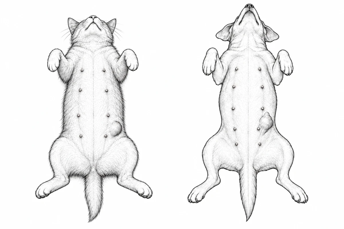
Mammary glands are the breast glands that make milk. They lie in two rows along the underside of a dog or cat. A mammary lump may involve one gland or several. A lump near a nipple is not always a mammary tumour, so an examination is needed to determine where it starts.
A tumour is an abnormal growth of cells. Some mammary tumours in dogs are benign, meaning they do not spread to other parts of the body. Others are cancerous and can grow into nearby tissue or spread. In cats, most mammary tumours are cancerous, so even a small lump needs prompt assessment.
Several lumps do not necessarily mean that one tumour has spread. In dogs, different lumps can be different types of tumour. The team measures and records each lump separately. Male dogs and cats can also develop mammary tumours.
Signs and symptoms
What you might notice
You may feel a small firm lump beside a nipple, or see a larger swelling. Some lumps move under the skin. Others feel attached to deeper tissue, grow quickly, or develop an open, bleeding surface. Your pet may lick the area or seem sore.
Tell your vet when you first noticed the change, how quickly it has grown, and whether there are other lumps. Mention previous surgery, hormone medicines, whether your pet is spayed (has had the ovaries removed), and any loss of appetite, weight loss or breathing changes.
Arrange urgent veterinary assessment for a rapidly swollen, hot, red or painful area involving several glands, especially if your pet seems unwell. Infection and a form of cancer called inflammatory mammary carcinoma can cause this pattern. It needs a different treatment plan from a single breast lump.
Diagnosis
Finding out what the lump is
Your vet will check every gland and measure each lump. A fine-needle aspirate, or FNA, collects cells through a small needle. It can identify some other kinds of lump, but it often cannot tell whether a mammary tumour is benign or cancerous.
That usually needs tissue examined in a laboratory. A biopsy removes a piece for testing; sometimes the whole lump is removed and tested instead. The choice depends on the lump’s size and location, and whether the result would change surgery.
Checking whether cancer has spread
Chest X-rays help check the lungs before surgery. Some pets need a more detailed scan, called a CT scan. An ultrasound of the abdomen may also help in selected cases; it is not needed for every pet. For cats, the surgical team decides about abdominal ultrasound after referral.
Lymph nodes are small structures that filter fluid draining from tissues. They can be a site of cancer spread. Dr. Wood plans which nodes need testing or removal. Sometimes mapping helps find which nodes receive fluid from the tumour area.
Treatment
Planning how much tissue to remove
In dogs, surgery may remove a lump, a gland, or a larger area. The number and position of the lumps, their size and attachment, and the skin available to close the wound all matter.
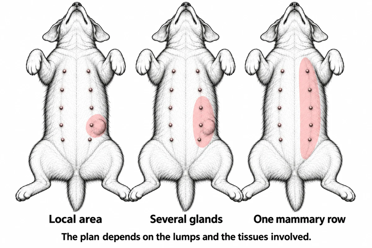
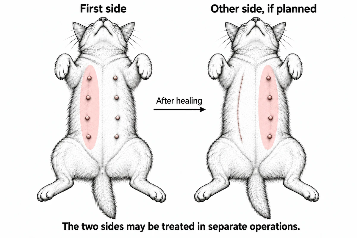
Cats usually need a larger operation when mammary cancer is suspected or confirmed. This may mean removing a whole row of glands on one or both sides. When both sides need treatment, the surgical team often plans separate operations so the first wound can heal before the second.
Inflammatory mammary carcinoma is different: surgery is not recommended to control that cancer. The plan focuses on pain relief and discussion of medical treatment with a cancer care team.
If your pet is not spayed, ask the surgical team whether spaying can be part of the plan. Spaying does not replace tumour treatment or guarantee that an existing cancer will behave better.
Before booking surgery, ask the surgical team about the expected hospital stay, help your pet may need at home and planned rechecks.
Aftercare and outcome
Healing and reviewing the results
Follow your pet’s discharge plan for pain medicine, activity, wound protection and rechecks. Prevent licking and check the wound each day. Contact the hospital if it opens, develops discharge or becomes more painful. The shared recovery guide has practical advice; your pet’s own instructions take priority.
Use the contact details on your discharge sheet. If you do not have them, contact your treating veterinary team. For an urgent or worsening concern, call rather than waiting for an email reply.
The tissue report identifies each tumour and checks the edges of the removed tissue, called the margins. It may also describe grade—features that help indicate how the cancer may behave—and whether cells have entered small blood vessels or fluid channels.
The tissue, lymph-node and scan results guide follow-up. Some dogs need monitoring; others need more surgery or a cancer specialist. Cats often need closer checks because their tumours are more likely to spread.
For veterinary teams Mammary mass referral checklist
← Back to the owner explanation
Mammary / breast lump: referral checklist
Before referral
- CBC / chemistry
- 3-view chest radiographs
- Mammary map: gland, size of each mass, fixation/ulceration, nodes and photos
- Growth, spay/hormone history, prior surgery and systemic signs
- Mass FNA if diagnosis unclear — include cytology/pathology report
Send: blood results, histopathology, original chest images/reports and previous op notes.
Coordinate with the surgical team
- Biopsy if it changes surgery, especially in cats or atypical masses
- Dr Wood plans node removal / sentinel-node mapping with mammary tumour surgery; no routine node FNA
- Feline abdominal US: case by case after referral. CT/abdominal staging for advanced disease or abnormal findings
Diffuse painful inflammation, rapid progression or systemic illness: urgent assessment and medical/oncology discussion.
Prepare a referral →Planning notes and references
AAHA diagnostic and staging guidance
Planning mammary surgery
Cats
Feline mammary tumours are more often malignant. When both chains require surgery, Dr Wood typically recommends staged bilateral mammary-chain surgery. The timing is planned from disease distribution, patient health, and closure requirements.
Dogs
The number, size, location, fixation and lymphatic drainage help determine whether to remove a limited area, a region or a whole mammary chain.
Staging depends on the species, examination and tumour findings. Arrange chest imaging before referral and send any existing images with their reports. For cats, the surgical team also considers regional lymph-node assessment. Abdominal ultrasound is selected case by case and arranged after referral. In dogs, abdominal imaging is selected when findings could change the plan. Dr Wood will plan lymph-node removal when needed and arrange sentinel lymph-node mapping before surgery when appropriate. AAHA 2026 species-specific staging guidance.
3 cm changes the prognosis discussion
Maximum tumour diameter is one of the simplest useful risk features to record before referral. It does not replace histology, but it helps frame staging and surgical urgency.
current mammary assessment guidance · feline mammary carcinoma study
 05Toe or nail-bed lesionA sore toe, swelling or a damaged nail
Overview
A sore toe is not always an infection
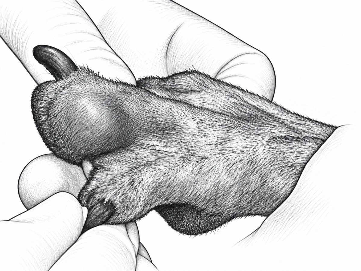
A swollen toe, damaged nail or sore nail bed can have several causes. Infection, injury, inflammation and tumours can look similar. The nail bed is the tissue where the nail grows. You may see a toe called a digit in your pet’s records.
Some toe tumours are benign, meaning they do not spread elsewhere. Others are cancerous and can grow into nearby tissue or spread. A persistent problem needs a diagnosis rather than repeated treatment based on its appearance.
The assessment looks for the cause of the change and checks whether the bone is affected. Dogs and cats need different assessments. A toe tumour does not automatically mean removing the whole leg.
Signs and symptoms
Changes worth checking
You may notice limping, licking, swelling, bleeding or discharge around a nail. A nail may break, fall off or grow in an unusual direction. Sometimes the only sign is a toe that stays sore or a wound that does not heal.
Tell your vet how long it has been there, whether treatment helped, and whether more than one toe is affected. Infection can occur alongside a tumour, so improvement with antibiotics does not always explain the whole problem.
Contact your vet promptly if your pet cannot comfortably use the foot, has worsening pain or swelling, or has a wound that keeps bleeding. Breathing difficulty needs emergency care.
Diagnosis
Checking the toe and getting a sample
Your vet will examine the foot and check the other toes. Foot X-rays show whether the bone is damaged and help plan treatment. Bone damage raises concern, but it does not prove cancer: infection and some benign conditions can also damage bone.
A fine-needle aspirate, or FNA, collects cells through a small needle. It can help with some tumours, but toe samples are often unclear. A biopsy removes tissue for laboratory testing. The team plans where and how to take the sample so that it does not make later surgery harder.
Why check beyond the foot?
Chest X-rays help check the lungs before a decision on surgery for a toe or nail-bed mass. Lymph nodes—small structures that filter fluid from tissues—may also need assessment because some cancers spread there. The tests depend on the suspected tumour and your pet’s examination.
For selected malignant toe tumours, such as melanoma, sentinel lymph node mapping can help find which node to check.
In cats, a toe problem can sometimes be caused by cancer that started in the lungs and spread to the toes. This is called lung-digit syndrome. It is particularly concerning when several toes or the bone are affected. Chest imaging is an important part of this assessment, even if your cat has no cough or other obvious breathing problem.
Treatment
When is a toe removed?
An infection or inflammatory problem needs treatment for that cause. If a tumour is suspected or confirmed, removing the affected toe may be the best way to remove it and relieve pain. Sometimes that operation also provides the tissue needed for a final diagnosis.
The operation depends on which toe is involved, how far the problem extends into bone and nearby tissue, and the condition of the rest of the foot. A tumour extending beyond one toe may need a different plan. Discuss expected walking ability and wound care with the surgical team before surgery.
Planning removal and wound closure →
If tests show disease elsewhere, the team considers whether surgery would improve comfort and what other treatment could help. A toe operation cannot cure cancer that has spread from the lungs. Further cancer treatment is based on the diagnosis, rather than given to every pet with a toe tumour.
Before booking surgery, ask the surgical team about the expected hospital stay, help your pet may need at home and planned rechecks.
Aftercare and outcome
Protecting the foot while it heals
Follow the discharge plan for pain medicine, short controlled walks, wound protection and rechecks. If a bandage is used, keep it clean and dry and follow the change schedule. Call if it slips, gets wet, smells, or your pet becomes more uncomfortable. Prevent licking or chewing.
Use the contact details on your discharge sheet. If you do not have them, contact your treating veterinary team. For an urgent or worsening concern, call rather than waiting for an email reply.
Many dogs walk well after one toe is removed, but healing and function depend on the toe, the wound and any other foot or joint problems. The surgical team reviews the tissue report to identify the tumour and check the edges of the removed tissue, called the margins.
The outlook depends on the tumour type and whether it has spread. Some toe cancers are controlled well with surgery; others need a discussion with a cancer specialist and closer monitoring. The shared recovery guide explains general home care; follow the foot and bandage instructions above and your own discharge plan.
For veterinary teams Toe or nail-bed lesion referral checklist
← Back to the owner explanation
Toe or nail-bed lesion: referral checklist
Before referral
- Lesion FNA if feasible — include cytology/pathology report
- CBC / chemistry
- Orthogonal digit/foot radiographs
- 3-view chest radiographs for digit/nail-bed masses; cats: check for pulmonary primary
- Digit(s), size, nail changes, pain/lameness, discharge, treatment response, nodes and photos
Send: blood results, biopsy reports, original foot/chest images and reports.
Coordinate with the surgical team
- Biopsy / planned digit amputation if FNA non-diagnostic
- MCT/STS of digit/web: tumour-specific staging
- Node assessment / sentinel-node mapping as part of toe / nail-bed tumour surgery; thoracic CT for high metastatic concern or abnormal films; local CT for proximal extension
Avoid unplanned excision if margins may be needed.
Prepare a referral →Planning notes and references
AAHA diagnostic and staging guidance · Feline digital carcinoma study
Planning toe surgery
Dogs
Persistent nail-bed lesions, especially with bone lysis, require a tumour work-up even when infection is also present. Radiographs and tissue diagnosis guide biopsy, digit amputation, and regional-node assessment.
Cats
A destructive digital lesion may be a primary tumour or metastatic disease. Thoracic imaging is particularly important because lung tumours can spread to the digits.
24 of 29 masses with bone loss were malignant
In a study of 117 dogs with 124 digit masses, 24 of the 29 masses with radiographic bone loss (83%) were malignant. This is the bone-loss subgroup, not all digit masses. Infection can still coexist, so drainage or inflammation should not end the tumour work-up.
 06Anal sac massA lump inside an anal sac
This guide is mainly about anal sac cancer in dogs. Anal sac tumours are rare in cats, and their treatment and outlook need a separate discussion.
Overview
Where are the anal sacs?
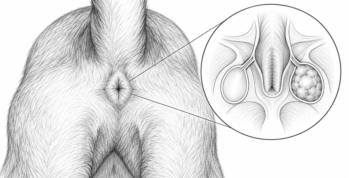
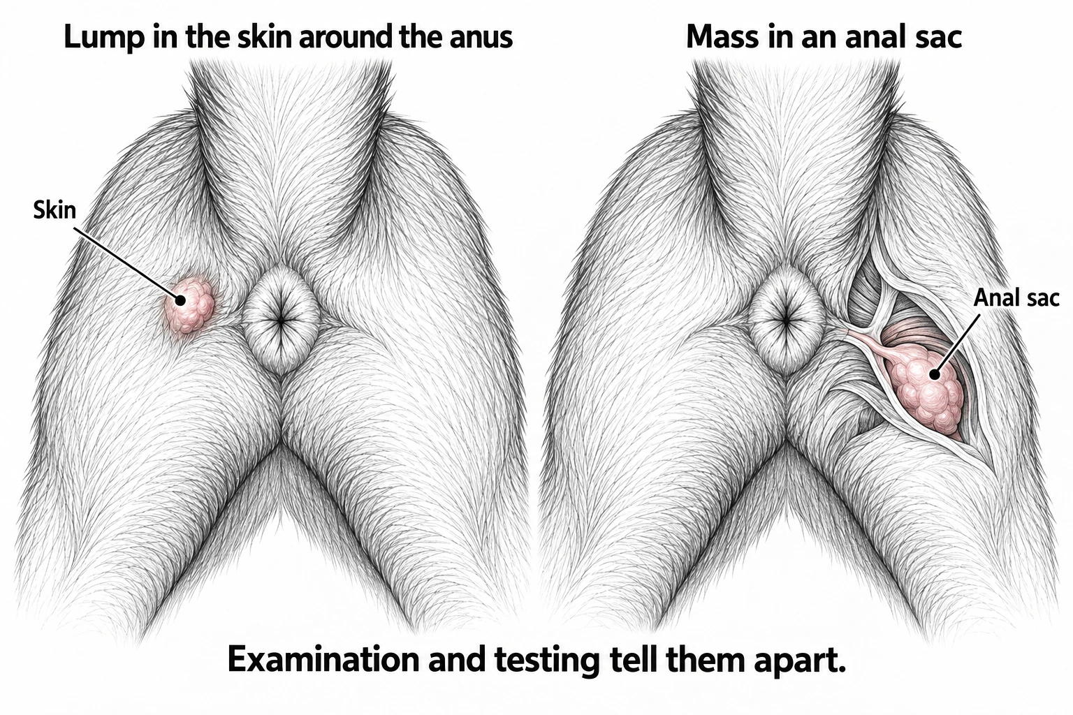
The anus is the opening where stool leaves the body. Just inside it are two small pouches, one on each side, called anal sacs. They hold a strong-smelling fluid. A lump within a sac differs from one in the surrounding skin.
In dogs, an anal sac mass may be a cancer called anal sac adenocarcinoma, shortened to AGASACA in reports. Infection and inflammation can also cause swelling. Examination and tests help tell them apart.
Why does it need checking?
Anal sac cancer can grow into nearby tissues or spread elsewhere. Even a small lump needs assessment; its size alone cannot show whether it has spread.
Signs and symptoms
What you might notice
You may see swelling beside the anus, licking, blood near the stool, or straining to pass stool. Some dogs have no obvious signs. Their vet finds the lump while examining the anal sacs.
Some of these tumours raise the calcium level in the blood. This can cause more drinking and urination, vomiting, poor appetite or weakness, and can affect the kidneys. These changes have other possible causes too; a blood test helps clarify the cause.
Tell your vet about trouble passing stool or urine, weight loss, discomfort, and changes in drinking. If your dog cannot pass urine, repeatedly strains without passing stool, or becomes very weak or unwell, contact a vet urgently.
Diagnosis
Examining and sampling the lump
Your vet examines both sacs and feels just inside the anus. This helps assess the lump's size, whether it moves freely, and whether anything is narrowing the passage for stool.
A fine-needle aspirate, or FNA, collects cells through a small needle. Looking at the cells under a microscope can help distinguish cancer from inflammation. After removal, the laboratory examines the tissue for a fuller diagnosis.
Checking the blood and looking for spread
Blood tests check general health, kidney function and calcium. The surgical team recommends a test called ionized calcium, which measures the active form of calcium in the blood. A high result may need treatment before an anaesthetic.
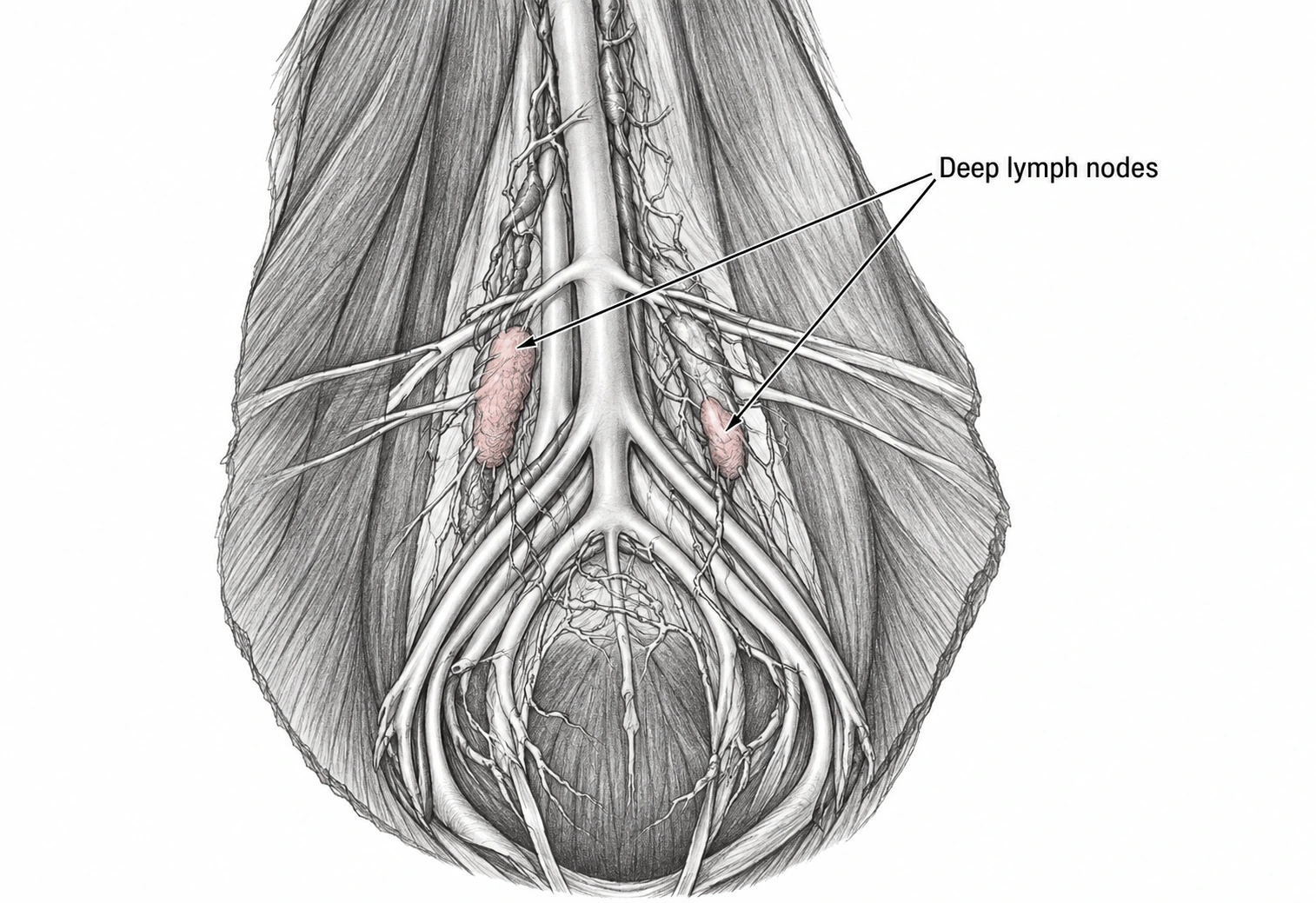
Lymph nodes are small structures that filter fluid from tissues. Anal sac cancer can spread to nodes deep in the back of the abdomen or inside the pelvis, the bony area around the hips. Enlarged nodes can press on the bowel or urinary tract. Assessment includes this area even when the original lump is small.
In selected dogs, sentinel lymph node mapping helps identify drainage for the node assessment. It is considered alongside the abdominal and pelvic imaging plan.
Chest X-rays check the lungs. Ultrasound or CT helps assess the abdomen and pelvis. CT uses X-rays to create detailed pictures, including pelvic nodes that are harder to assess by ultrasound. After referral, the surgical team uses the examination, blood results, signs and planned treatment to decide whether CT would help.
Scans help with planning, but a node's size alone cannot prove or rule out cancer. A cell or tissue sample may be needed. What samples and scans can show →
Treatment
Planning surgery
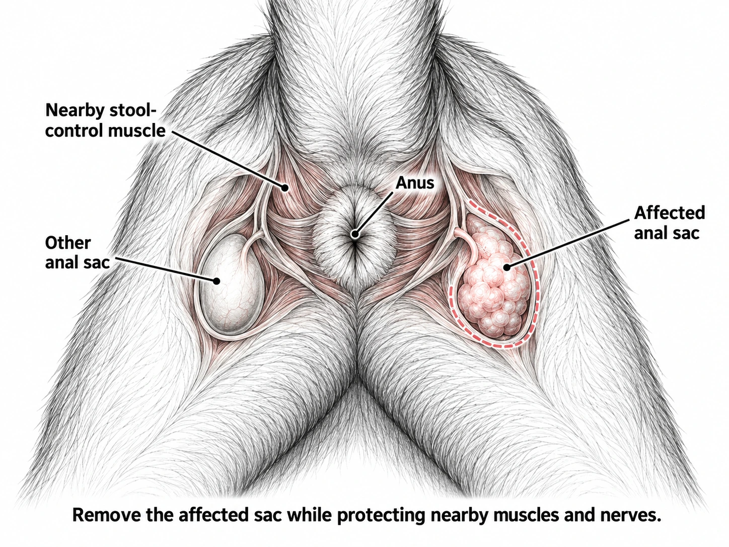
For a dog with suspected or confirmed anal sac adenocarcinoma (AGASACA), Dr. Wood recommends removing both anal sacs when surgery is suitable. The opposite sac can contain cancer even when it looks and feels normal. During surgery, Dr. Wood removes the tumour and both sacs while protecting the nearby muscles and nerves that control stool. Both sacs go to the laboratory for examination.
A 2024 study found hidden cancer in the opposite sac in 7 of 35 dogs whose work-up suggested cancer on only one side. This finding supports Dr. Wood’s recommendation. It does not prove a survival benefit or that one-sided and two-sided surgery carry equal risks. Read the study.
This recommendation concerns suitable dogs with this suspected or confirmed cancer. Benign anal-sac swellings, lumps in the surrounding skin and anal-sac disease in cats need their own assessment. Discuss your dog’s surgical risks and recovery with the surgical team before proceeding. How the operation and wound closure are planned →
If cancer has reached lymph nodes, removing the anal sac alone leaves disease behind. Selected nodes may also be removed through an operation in the abdomen. Their location, nearby blood vessels and your dog's health affect what is possible. Spread to a node does not automatically rule out treatment.
The surgical team may discuss radiation to treat disease in a chosen area, or cancer medicines with a cancer care team. These are not needed for every dog. Their likely benefit and side effects depend on the findings. If surgery is unsuitable, ask the surgical team about other ways to control the disease and keep your dog comfortable.
Before booking surgery, ask the surgical team about the expected hospital stay, help your pet may need at home and planned rechecks.
Aftercare and outcome
Passing stool and protecting the wound
Follow the discharge instructions and general recovery guidance for pain relief, activity, wound protection and rechecks. Prevent licking with the protection provided. Give stool-softening medicine only if prescribed. If stool gets near the wound, follow the cleaning and contact guidance.
Watch how easily your dog passes stool and urine. Call if the wound opens, develops discharge or becomes more painful, or if your dog struggles to pass stool. Some dogs have trouble controlling stool after surgery. This may be temporary, but lasting problems are possible; the risk depends on the tumour and the operation.
Use the contact details on your discharge sheet. If you do not have them, contact your treating veterinary team. For an urgent or worsening concern, call rather than waiting for an email reply.
What happens next?
The surgical team reviews the tissue and any lymph-node results with you. The outlook depends on the tumour, spread, calcium level and treatment. Some dogs live well for a long time after treatment; the outlook varies.
Follow-up may include examination of both sac areas, blood tests and imaging. Cancer can return in the original area or in lymph nodes, so feeling well does not replace planned checks.
For veterinary teams Anal sac mass referral checklist
Anal sac mass: referral checklist
Before referral
- Mass FNA if feasible — include cytology/pathology report. Do not delay classic anal sac mass referral for FNA.
- CBC / chemistry, renal values and ionized calcium.
- 3-view chest radiographs.
- Rectal exam: both sacs, side, size, fixation and pelvic mass effect.
- Stool/urinary signs, PU/PD, pain, weight loss and prior treatment.
Send: histopathology, blood results, original images/reports and clinical notes.
Coordinate with the surgical team
- Abdominal/pelvic staging; CT case by case after referral, considering mass >2.5 cm or clinical/nodal concerns.
- Node assessment / sampling; sentinel-node mapping by the surgical service; hypercalcaemia treatment if indicated.
If ionized calcium is unavailable, the surgical team can arrange it. Assess regional disease even with a small primary. Feline cases: discuss separately.
Prepare a referral →Planning notes and references
AAHA diagnostic and staging guidance
Planning anal-sac surgery
CT planning: selected after referral
CT of the abdomen and pelvis is selected case by case after referral. A primary mass >2.5 cm is one planning flag, considered alongside the other preoperative findings.
Preoperative findings that may favour CT
- Primary mass >2.5 cm
- Hypercalcemia
- Tenesmus, dysuria or constipation
- A palpable sublumbar or pelvic mass
- Pelvic pain, lameness or bone pain
- Weight loss
- Definitive surgical treatment being considered
These findings guide the individual plan; the size flag is not a requirement for CT, and a small primary does not exclude regional disease.
The historical clinical scheme separates primary tumours at 2.5 cm. However, a small primary mass or normal calcium does not rule out regional nodal disease.
How CT findings are interpreted
- In one series, 20% of dogs with a primary mass under 2 cm already had confirmed lymph-node metastasis.
- One bilateral-surgery study treated a regional node over 5 mm, or abnormal in shape or internal appearance, as suspicious. Size alone does not confirm metastasis.
- Cytology or histopathology is used to establish nodal metastasis. Once nodal disease is confirmed, the published clinical scheme separates nodal stages at 4.5 cm.
- CT also shows the relationship to the rectum, pelvic canal, vessels, and ureters when surgery is planned.
small-primary metastasis study · bilateral anal-sac study and staging scheme
20% had hidden bilateral disease
In a retrospective series, histopathology identified occult contralateral AGASACA in 7/35 dogs with apparently unilateral disease. Dr Wood recommends bilateral anal sacculectomy for suitable dogs with suspected or confirmed AGASACA. This series had no concurrent unilateral control group and does not establish a survival benefit or equivalent complication risk.
These totals include normal nodes. CT identified more normal nodes, but not more abnormal nodes in this comparison; these are not counts of metastatic nodes.
bilateral anal-sac study · CT versus ultrasound study · CT staging study
 07Lump around the anusA lump in the skin around the anus
This guide is about lumps in the skin or glands around a dog's anus. A lump within an anal sac follows a different assessment. Cats with a lump in this area need a separate discussion.
Overview
Not every lump here is the same
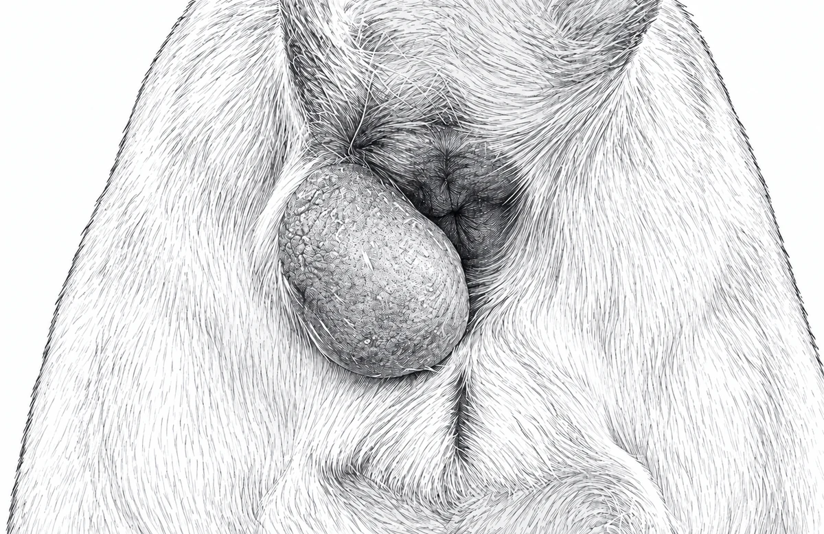
The anus is the opening where stool leaves the body. The skin around it contains several kinds of glands and other tissues. A lump may grow from any of them. The anal sacs are two pouches just inside the anus; a tumour within one of those sacs is a different problem.
Some skin-gland lumps are benign, meaning they do not spread elsewhere. Others are malignant, meaning they can grow into nearby tissues or spread. The surface appearance cannot reliably distinguish the two.
Example of a lump in the surrounding skin
One common benign type in male dogs that have not been neutered is a perianal adenoma. It grows from skin glands and can respond to male hormones. A cancer from those glands is called an adenocarcinoma. Check which diagnosis the report gives.
Signs and symptoms
Changes to look for
You may notice one lump or several, licking, irritation, bleeding or a sore surface. A larger lump may make passing stool uncomfortable. A small lump may cause no obvious problem at first.
Tell your vet when it appeared, how quickly it has grown, whether it has bled, and whether a lump was removed from that spot before. Mention changes in passing stool or urine, appetite, weight or energy.
Bleeding or rapid growth deserves assessment, but neither proves cancer on its own. Seek urgent veterinary advice if your dog cannot pass urine, repeatedly strains without passing stool, or has heavy bleeding or marked pain.
Diagnosis
Finding where the lump starts
Your vet examines the skin, feels just inside the anus and checks both anal sacs. The examination records the lump's size, whether it moves freely and whether it narrows the opening. Your dog's sex and neutering history are useful clues, but they do not establish the diagnosis.
A fine-needle aspirate, or FNA, collects cells with a small needle for examination under a microscope. For some gland lumps, the cells do not clearly show whether the growth is benign or malignant. A biopsy removes a piece of tissue and may be needed before choosing the operation.
Does every dog need scans?
No. A typical benign gland lump may not need tests for spread. If cancer is suspected, chest X-rays and selected abdominal scans help assess its extent.
Lymph nodes are small structures that filter fluid draining from tissues. The surgical team may assess nodes deep in the abdomen or pelvis—the bony area around the hips—when there is concern about spread. A CT scan gives detailed pictures of a deep or fixed lump and nearby structures. It can help plan a difficult operation, especially if a lump has returned or affects the passage for stool.
Sampling first can change how much tissue needs removal and how the wound can be closed. Avoiding an unplanned first removal can make later treatment simpler.
Treatment
When does surgery help?
For a benign, hormone-responsive gland tumour in a male dog that has not been neutered, neutering (removing the testicles) may help control the growth. Some lumps also need removal, particularly if they are large, sore or bleeding. Neutering is not a treatment for every lump in this area and is not a cure for a malignant tumour.
For cancer, the surgical team plans removal of the lump with some surrounding tissue when possible. The anus and the muscles that control stool are close by. The amount removed must balance control of the tumour with wound healing and bowel function. Larger or deeper wounds may need nearby skin moved to close them.
The laboratory examines the removed tissue and its edges, called margins. If tumour cells reach an edge, cells may remain in the wound. The surgical team uses this report and any tests for spread to discuss monitoring, further surgery or treatment with a cancer care team.
Before booking surgery, ask the surgical team about the expected hospital stay, help your pet may need at home and planned rechecks.
Aftercare and outcome
Keeping the wound protected
Follow your dog's written instructions for activity, medicines and rechecks; the shared recovery guide explains general care. Prevent licking with the protection recommended by the surgery team. If stool gets near the wound, use the cleaning and contact guidance. Do not put creams or other products on it unless prescribed.
Watch for increasing pain, swelling, discharge, bleeding or an open wound. Call if your dog has trouble passing stool. Operations close to the anus can sometimes affect stool control or leave a narrower opening. Discuss these risks with the surgical team in relation to the operation your dog needs.
Use the contact details on your discharge sheet. If you do not have them, contact your treating veterinary team. For an urgent or worsening concern, call rather than waiting for an email reply.
Reviewing the results
The outlook depends on what the lump is, whether it was fully removed and whether it has spread. A benign gland lump and a malignant tumour need different follow-up. Check the area for a new lump or regrowth, and keep the planned rechecks even if your dog seems comfortable.
For veterinary teams Lump around the anus referral checklist
Lump around the anus: referral checklist
Before referral
- Mass FNA if diagnosis unclear — include cytology/pathology report.
- Rectal exam: both anal sacs; mass site, number, size, fixation, ulceration and photos.
- Neuter status, growth, recurrence/prior surgery and stool/urinary signs.
- CBC / chemistry and calcium if malignancy suspected.
- Chest imaging: if malignancy suspected; not routine for classic benign adenoma.
Send: histopathology, blood results and original images/reports.
Coordinate with the surgical team
- Biopsy if malignancy possible; plan site before excision.
- Abdominal/pelvic staging and CT for invasive, fixed, recurrent or obstructive disease.
Suspected hepatoid adenocarcinoma >5 cm: additional CT planning flag.
Prepare a referral →Planning notes and references
AAHA diagnostic and staging guidance
Planning removal of a superficial mass
A small, superficial perianal-gland mass in a typical patient may need only FNA, rectal examination, and a simple-removal plan.
When CT or biopsy may be added
They may be recommended when a suspected perianal or hepatoid-gland tumour is over 5 cm, fixed or invasive, recurrent, of uncertain tissue origin, or when the result could change the margins or closure plan. The 5 cm finding does not apply to anal-sac cancer.
Tumour size and origin
Size, fixation, invasion and tissue of origin inform staging and surgical planning. Evidence about perianal or hepatoid-gland adenocarcinoma comes from selected tumour cohorts; it does not describe every lump around the anus.
This evidence does not apply to anal-sac apocrine gland adenocarcinoma, which has a separate pathway above. Review the hepatoid-gland tumour evidence.
Arrange an assessment
At the consultation, the surgical team will discuss treatment and recovery.
Send your vet’s notes, test results, images and any previous biopsy or surgery reports.