
Ischaemia-reperfusion injury
Read the draft explanation
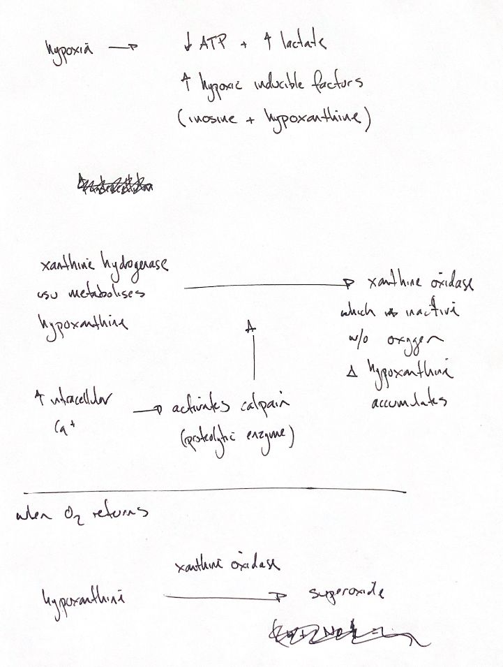
During ischaemia, reduced blood flow limits oxygen and fuel delivery. ATP falls and lactate can accumulate. ATP breakdown also leaves inosine and hypoxanthine. These are purine metabolites. They are not hypoxia-inducible factors, despite the way the original brackets them. HIFs are transcription factors that help cells respond to low oxygen.
The left-hand enzyme is xanthine dehydrogenase, or XDH, rather than “xanthine hydrogenase”. It normally oxidises hypoxanthine. The classic drawing links increased intracellular calcium to calpain activation, then XDH conversion to xanthine oxidase, or XO. Calpain is a protease: it cuts proteins.
That calpain link needs a dashed arrow. In a purified rat-liver enzyme experiment, calpain I and II did not convert XDH to XO. Conversion also did not increase in one early canine cerebral-ischaemia experiment. The pathway depends on the organ, species and model; it is not a proven sequence in every hypoxic canine tissue. Other changes, including oxidation or limited proteolysis, can shift XDH toward XO.
During anoxia, oxygen-dependent XO turnover stalls and hypoxanthine can build up. When oxygen returns, XO can oxidise this purine toward xanthine. Electrons from that oxidation pass through the enzyme to oxygen. A one-electron reduction of O₂ forms superoxide, O₂•−. Hypoxanthine does not turn into an oxygen radical molecule.
XO also makes hydrogen peroxide, H₂O₂. Superoxide is therefore not its only reactive oxygen product. These oxidants can add to tissue injury during reperfusion. The sketch follows one source pathway; it does not account for every source of reperfusion damage.
References
Johnston & Tobias, Veterinary Surgery: Small Animal, 2nd ed., Ch. 6; Stark et al., Biochem Biophys Res Commun 1989;165:858–864; Mink et al., Am J Physiol 1990;259:H1655–H1659; Kelley et al., Free Radic Biol Med 2010;48:493–498; Wang & Semenza, J Biol Chem 1993;268:21513–21518.