
Type A and type B lactic acidosis; LDH and concurrent acid load
Read the draft explanation
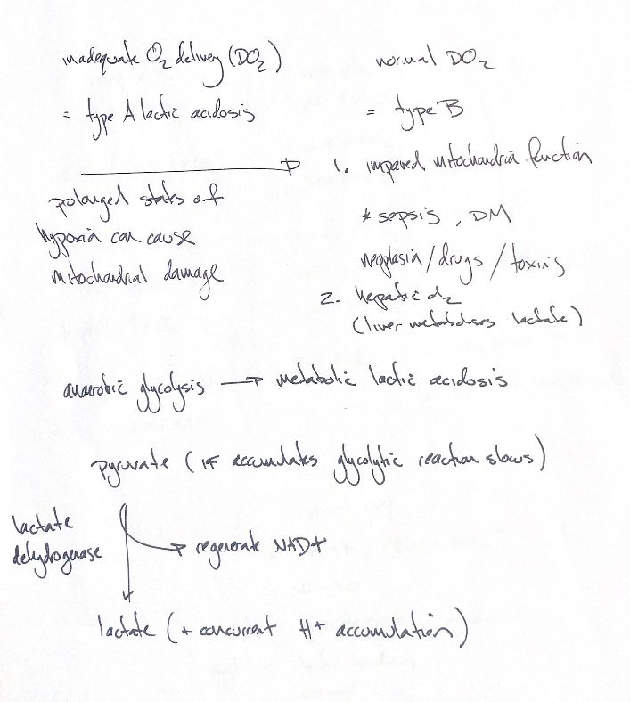
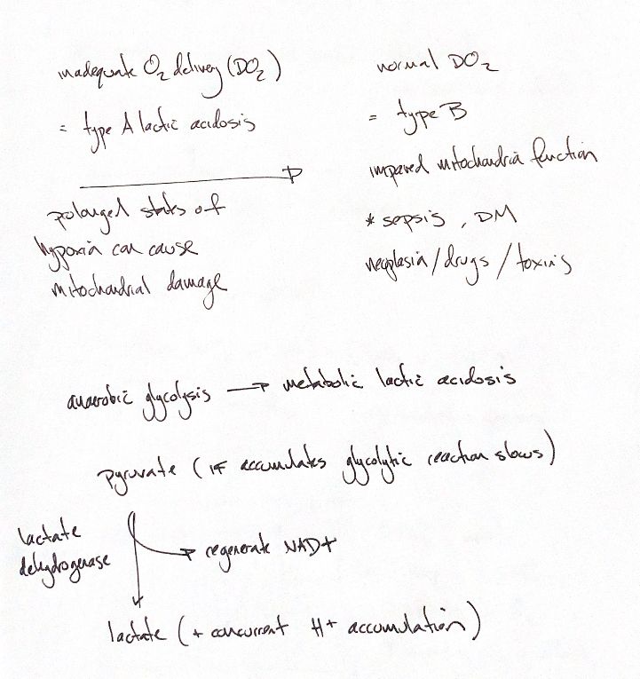
Type A and type B describe different routes to a high lactate result. The original calls them types of lactic acidosis. A high lactate value alone does not prove metabolic acidosis.
In type A, oxygen delivery, or DO₂, is inadequate. Cells rely more on anaerobic metabolism. In type B, lactate can rise despite adequate oxygen delivery. Impaired mitochondrial function is one mechanism. The original examples are sepsis, diabetes mellitus, neoplasia, drugs and toxins. These conditions can have more than one mechanism. Prolonged hypoxia can damage mitochondria, so type A and type B processes may overlap.
Keep the liver branch from the first drawing. The liver metabolises lactate. Hepatic disease can reduce clearance, letting blood lactate rise even without a rise in production. Blood lactate reflects both production and clearance.
Now follow glucose through glycolysis to pyruvate. The original links pyruvate build-up with slower glycolysis. The key step shown here is NAD⁺ recycling, rather than pyruvate accumulation alone. Lactate dehydrogenase, or LDH, converts pyruvate to lactate while regenerating NAD⁺. Recycling NAD⁺ allows glycolysis to continue.
The reaction is pyruvate + NADH + H⁺ → lactate + NAD⁺. In this direction, LDH uses H⁺ rather than releasing it. The source also shows concurrent H⁺ accumulation. Keep that as a separate process: during inadequate oxygen delivery, acid can accumulate when its production exceeds use. Metabolic acidosis may accompany rising lactate, but the two findings are not the same measurement.
References
Chris Wood’s two drawings; Johnston & Tobias, Veterinary Surgery: Small Animal, 2nd ed., ch. 6; IUBMB EC 1.1.1.27; Saint-Pierre, Epstein & Hopper 2026, DOI 10.1111/vec.70148.