
Healing tissue strength
Read the draft explanation
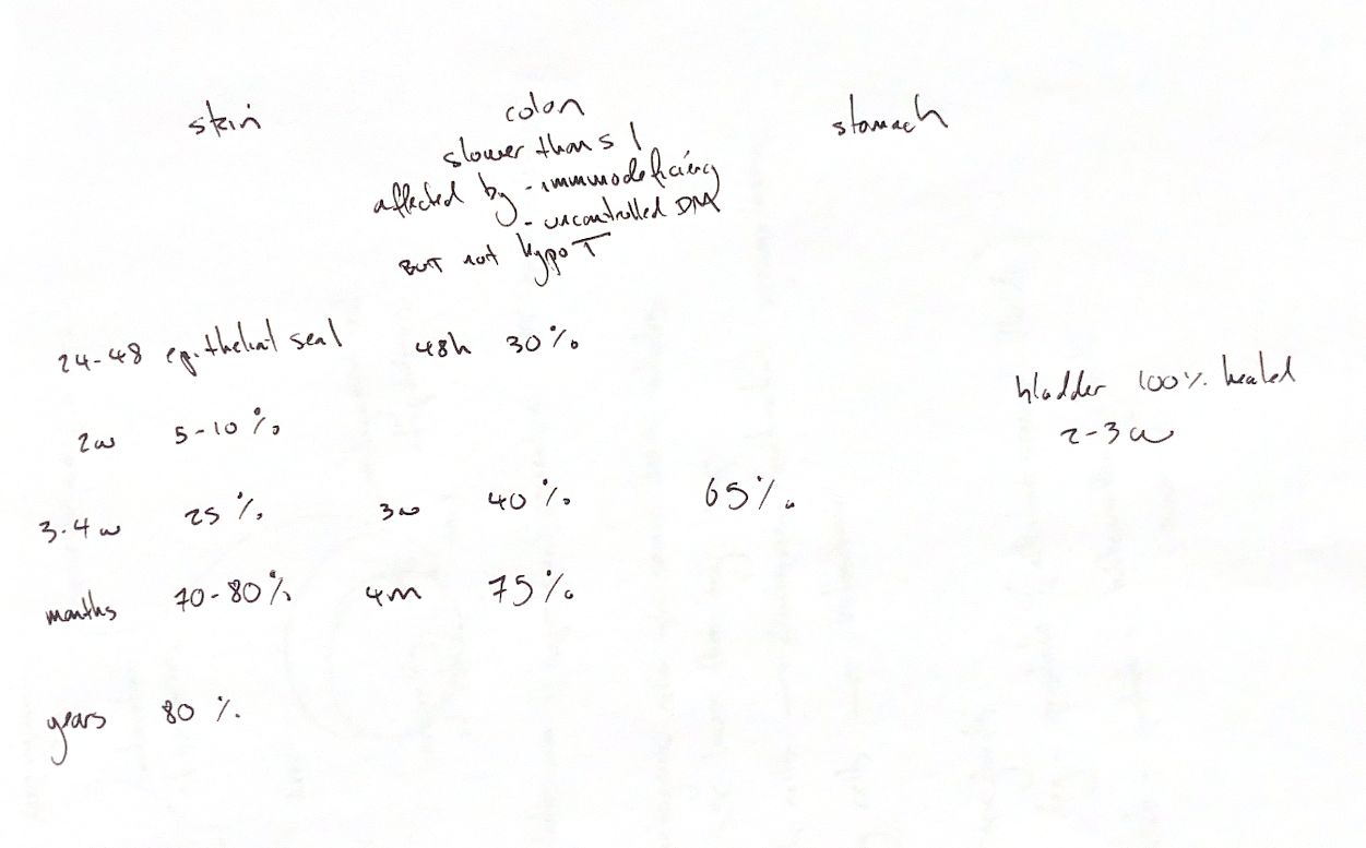
A closed surface can still cover a weak wound. Well-apposed skin may form an epithelial seal in 24–48 hours. Approximate strength landmarks are 5–10% at 2 weeks, 25% at 3–4 weeks and 70–80% after several months. The long-term 80% cue includes a one-year rat-incision example in the textbook, rather than a measured timeline for every dog. Collagen alignment and cross-linking strengthen the scar.
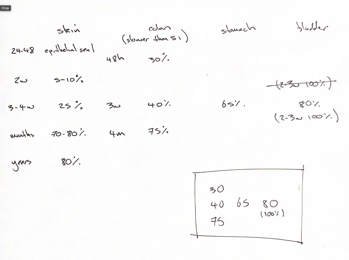
Read the GI figures as approximate study landmarks, not one measured dataset. The colon cues are 30% at 48 hours, 40% at 3 weeks and 75% at 4 months. Stomach is 65% at 3 weeks. Colonic strength returns more slowly than small-intestinal strength. Early collagen breakdown weakens the repair while sutures hold the edges.
The figures depend on species, model and strength test. The textbook repeats the 30% and 75% colon estimates, but its comparison graph differs from the prose. The exact 40% and 65% landmarks are not established by the accessible primary abstracts. The 1975 dog experiment's summary reports stomach and colon no stronger than 70% of normal at 120 days, which differs from the 75% teaching estimate. Bursting pressure and breaking strength are different tests.
Normal bladder defects can regain 100% of unwounded strength by about 3 weeks. The textbook gives 14–21 days; the primary experiment supports recovery within 21 days. Healing activity continues after strength returns. One original variant also shows 80% at 3–4 weeks, beside a crossed-out and then repeated 100% at 2–3 weeks. That 80% alternative conflicts with the matched bladder evidence. The boxed 30/40/65/80(100)/75 cue therefore needs its tissue, time and qualification attached.
Immunodeficiency and poorly controlled diabetes can impair repair. The original “not hypothyroid” cue is too broad. Clinical colonic effects remain uncertain in the cited textbook review, while hypothyroid rats in another experiment had weaker anastomoses and less collagen. Species and endpoint matter.
References
Johnston/Tobias, 2nd ed., Chs. 9, 75, 93, 116, 121; Hastings et al., Surg Gynecol Obstet 1975;140:701–707, 933–937; Thá Nassif et al., Eur Surg Res 2009;42:209–215.