
Intrinsic laryngeal muscles and glottic closure
Read the draft explanation
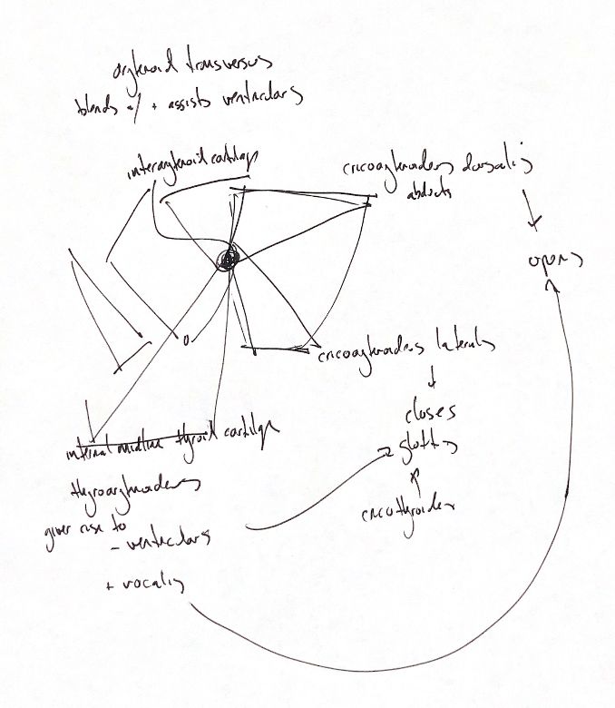
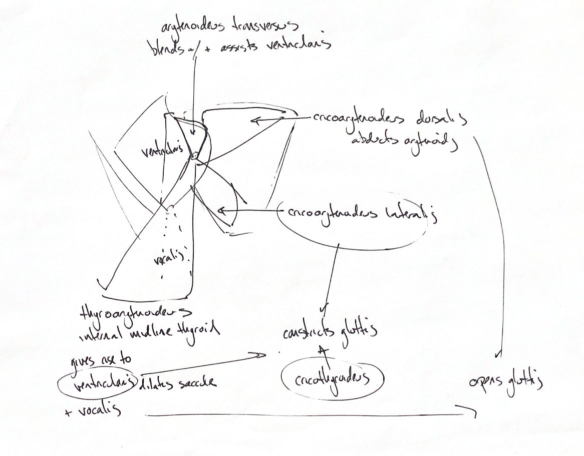
In the dog, cricoarytenoideus dorsalis abducts the arytenoid to open the glottis. Lateralis pivots it medially to close the rima glottidis. One handwritten variant calls dorsalis “adducting”; the checked opening action is abduction.
Both reach the arytenoid muscular process, from different cricoid surfaces. Thyroarytenoideus is the parent muscle mass for ventricularis and vocalis. That relationship does not give them identical adult attachments: canine ventricularis arises from the cuneiform process, while vocalis inserts on the vocal process.
Ventricularis constricts the glottis and dilates the laryngeal saccule. Vocalis draws the arytenoid ventrally and relaxes the vocal cord; the “opens” shorthand should not turn it into the primary abductor. Arytenoideus transversus reaches the interarytenoid cartilage and blends with ventricularis, assisting vocal-fold adduction and glottic constriction.
Cricothyroideus lies between the thyroid lamina and cricoid. It pivots the cricoid on its thyroid articulation to tense the vocal cords; its grouping beside “closes” in the sketch needs that distinction.
The small hyoepiglotticus muscles run from the ceratohyoid to a shared band on the ventral epiglottic midline and draw the epiglottis ventrally. Detailed muscle passages support that origin; broad epiglottic passages in both books instead describe a hyoid-body connection. Hyoepiglotticus receives hypoglossal (XII) supply, so the list is not a universal vagal-supply rule.
The reference views have different cuts, removals and reflections; absent muscles should not be mistaken for normal absence.
References
Johnston & Tobias, 2nd ed., Ch 101; Miller’s Anatomy of the Dog, 4th ed., Ch 6 and 8; Skerrett et al. 2015.