
Extrinsic laryngeal muscles
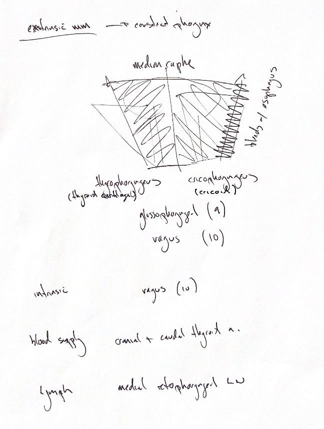
Read the draft explanation
The canine thyropharyngeus and cricopharyngeus help constrict the caudal pharynx. Both attach to the median raphe of the dorsal pharynx—the midline connective-tissue seam.
Thyropharyngeus is a large, flat muscle covering the outside of the larynx. It extends from the lateral surface of the thyroid cartilage to the dorsal pharyngeal raphe. Cricopharyngeus lies on the larynx and pharynx immediately caudal to it, extending from the lateral cricoid cartilage to the same raphe. Some fibres along its caudal margin blend with the oesophagus. Constriction is only part of the action: coordinated relaxation is also needed during swallowing.
The note and textbook group their innervation as glossopharyngeal (IX) and vagal (X). Canine experiments support contributions from both through the pharyngeal plexus, with variation between individuals. Cricopharyngeus motor supply is described more specifically through the pharyngo-oesophageal nerve, primarily from the vagus. The shared IX/X cue does not imply equal contributions to every muscle.
The separate intrinsic-muscle reminder is mainly vagal, through cranial and caudal laryngeal branches. Cricothyroideus receives the external cranial branch. This is not a rule for every associated muscle: hyoepiglotticus receives hypoglossal (XII) supply.
Laryngeal blood supply comes mainly through branches of the cranial and caudal thyroid arteries. Lymph drains to the medial retropharyngeal lymph nodes.
References
Johnston & Tobias, Veterinary Surgery: Small Animal, 2nd ed., Ch 101; Venker-van Haagen et al. 1986; Lynch et al. 1973; Hudson 1986; Skerrett et al. 2015.