
Laryngeal cartilages — source views, attachments and species differences
Read the draft explanation
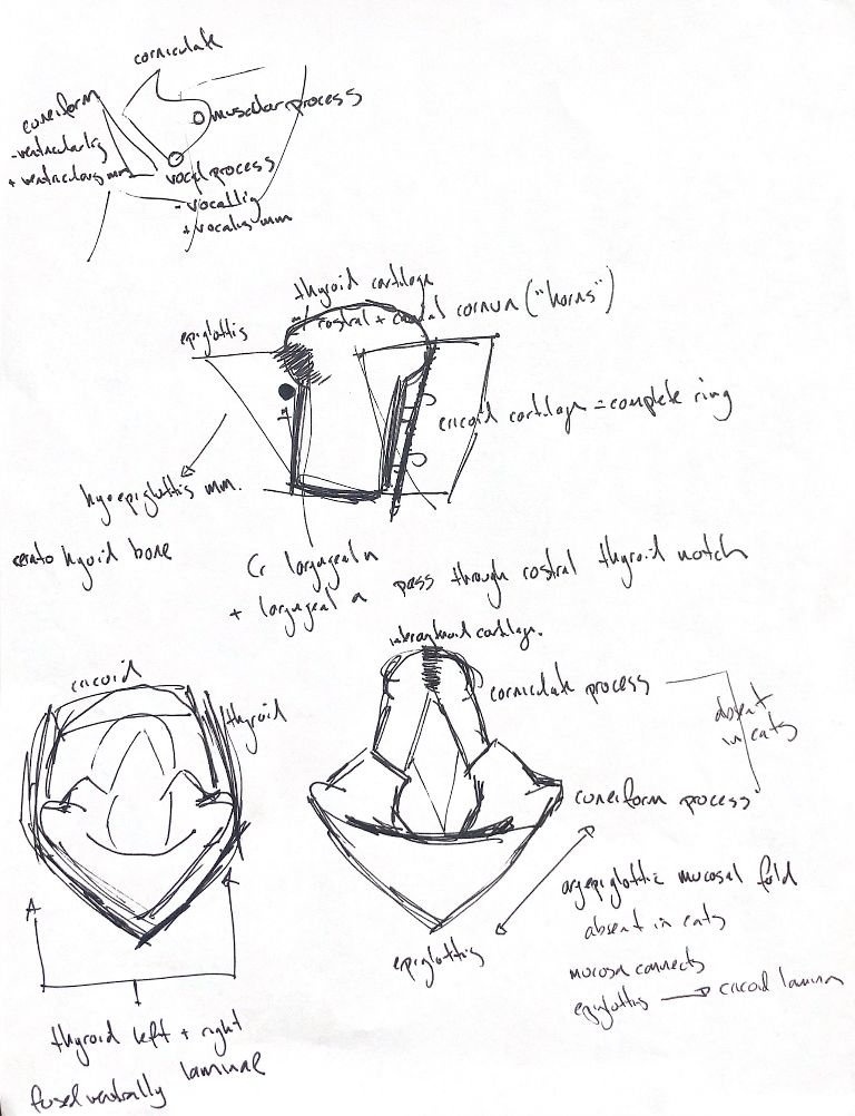
Start with the canine framework: epiglottic, thyroid, cricoid, sesamoid and interarytenoid cartilages, plus paired arytenoids. The thyroid laminae meet ventrally. The cricoid forms a complete ring. Each arytenoid has cuneiform, corniculate, muscular and vocal processes.
The epiglottis points rostrally and normally rests on the soft palate. Hyoepiglotticus draws it ventrally. The attachment descriptions differ: the typed note and gross-anatomy passages say the middle of the hyoid body. Miller’s detailed muscle passage and the later Johnston/Tobias passage describe the medial ceratohyoid origin. Miller describes a common tendon inserting on the ventral epiglottis. Both wordings remain visible.
Two Amis studies support active epiglottic control during breathing. One stimulated the muscle in four anaesthetised, tracheostomised, mechanically ventilated dogs. The other recorded breathing-related muscle activity in five anaesthetised, tracheostomised dogs. These are abstract-level experimental findings, not a promise about swallowing in an individual patient.
Follow the processes to their attachments. Ventricularis and its ligament attach at the cuneiform process. Vocalis and the vocal ligament attach at the vocal process. Keep another wording difference clear: the typed note places the ventral cuneiform part in the aryepiglottic fold; Miller specifies the vestibular fold and a dorsal aryepiglottic attachment.
Cats lack the canine cuneiform and corniculate processes and true aryepiglottic folds. Their mucosa connects the epiglottis to the cricoid lamina. In the transoral view, the patient’s left is on your right.
References
Johnston & Tobias, 2nd ed., Ch 101; Miller’s Anatomy of the Dog, 4th ed., selected muscle and larynx passages; Amis et al. 1996, two abstracts.