
Salivary gland openings and secretion
Read the draft explanation
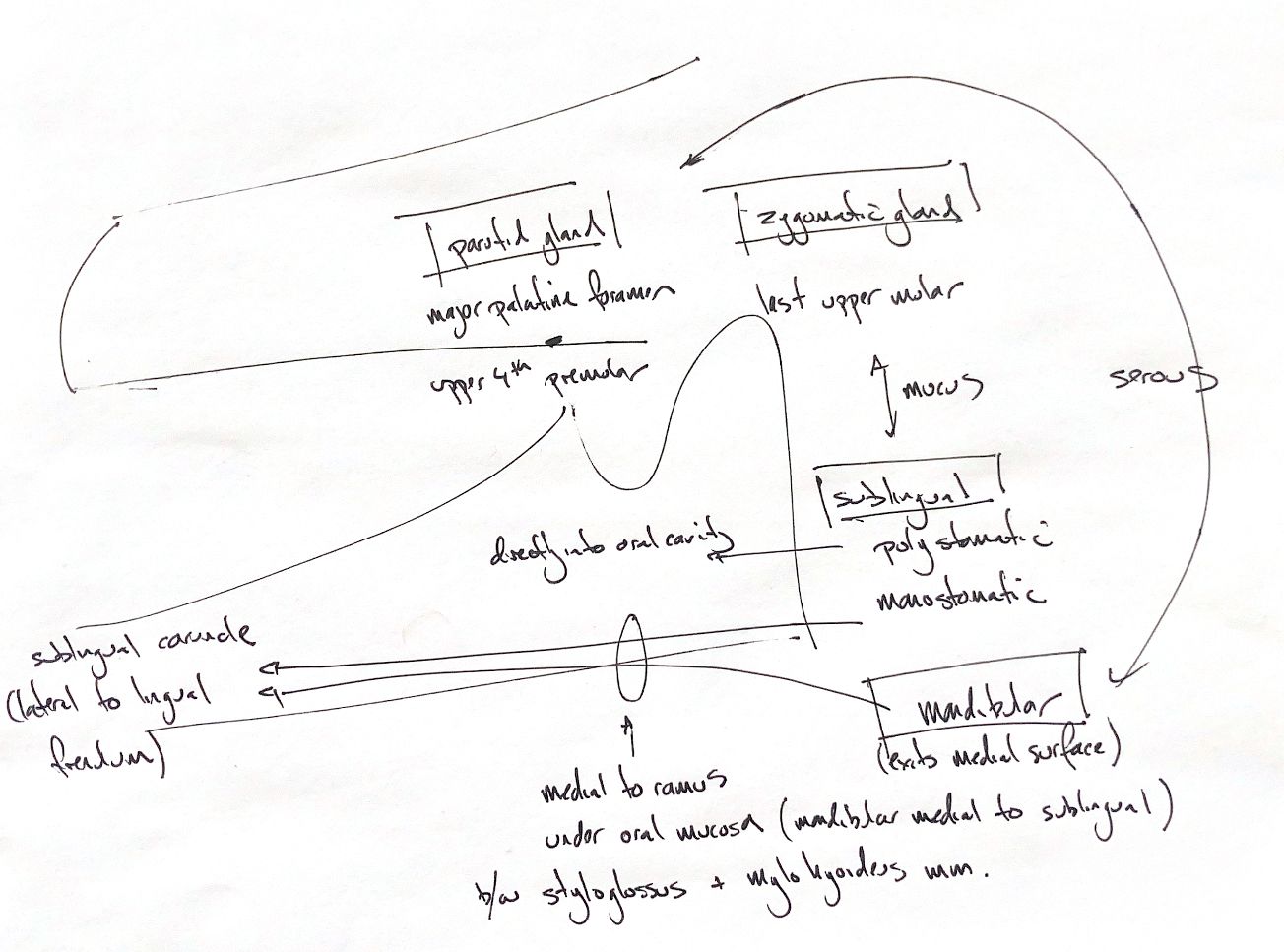
The parotid duct opens on a small papilla beside the upper fourth premolar. The major zygomatic duct opens caudolateral to the last upper molar. Both drain into the oral vestibule. The major palatine foramen is a separate hard-palate landmark, not a salivary duct opening.
The mandibular duct leaves the gland medially and runs rostrally, medial to the sublingual gland and mandibular ramus. Beneath the oral mucosa, it passes between styloglossus and mylohyoideus to reach the sublingual caruncle, lateral to the lingual frenulum.
The monostomatic sublingual duct runs beside the mandibular duct and usually opens at the caruncle just caudal to it. Polystomatic sublingual tissue has multiple small ducts opening directly into the oral cavity.
The serous/mucus comparison is a relative recall cue. Johnston & Tobias describes parotid and mandibular secretions as generally more serous, and sublingual and zygomatic secretions as richer in mucus. Most glands contain a mixture of secretory cells.
Canine histology adds an important limit: Gomi and colleagues found mucous-cell predominance in the mandibular and zygomatic glands, while monostomatic and polystomatic sublingual tissue differed. Cellular composition is not the same measurement as the character of the final saliva. Earlier puppy histochemistry also varied with developmental stage. These findings do not support a pure-serous/pure-mucous rule for every dog or cat.
References
Johnston & Tobias, 2nd ed., Ch 88; UMN carnivore anatomy, salivary glands; Gomi et al. 2017, abstract and selected histology results; Reifel & Travill 1972, publisher abstract.