
Alar folds, nasal movement and olfaction
Read the draft explanation
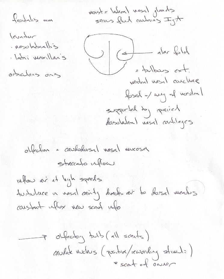
The alar fold is a bulbous rostral extension of the ventral nasal concha that joins the nostril wing. Paired dorsolateral nasal cartilages support the wings. In the frontal sketch, the patient's left stays on the viewer's right.
The muscle list recalls frontalis, levator nasolabialis, levator labii maxillaris and orbicularis oris. The book describes the levators and a branch of orbicularis oris widening the nares. Orbicularis oris also pulls the nose ventrally for downward sniffing. Frontalis needs a separate limit: the passage describes its action on the ear's scutiform cartilage, rather than a nasal-dilator action.
In dogs, paired lateral nasal glands supply much of the fluid that keeps the nasal planum moist. Their serous secretion contains IgA, or immunoglobulin A, suggesting a defensive role.
Olfactory receptors lie in the caudodorsal nasal mucosa. Rapid, staccato sniffs bring new scent samples through the dorsal meatus. The note's turbulence cue needs location: mixing can occur in the nasal vestibule, while a canine airflow model showed laminar flow through the olfactory recess. Odorants bind receptors on olfactory-cell cilia and start electrical signals in olfactory nerves.
The lower words link the olfactory bulb with scents and the caudate nucleus with positive or rewarding associations. The “owner scent” reminder is an attributed shorthand. Berns and colleagues studied 12 awake dogs with five odours. Bulb/peduncle activation was similar across those scents; the caudate responded most to the familiar human. That person was not the current handler and was absent during scanning. This suggests a positive association, not a direct measure of emotion or a universal test of owner preference.
References
Johnston & Tobias, 2nd ed., Ch99; Adams et al. 1981; Craven et al. 2010; Berns et al. 2015, abstracts and selected airflow text.