
Why action potentials normally travel forwards
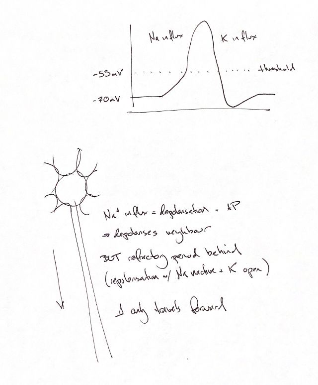
The falling phase in the original curve labels potassium influx. Potassium leaves the cell during repolarisation.
Read the draft explanation
An action potential, or AP, is a brief change in membrane voltage that carries a nerve signal. Na+ enters through voltage-gated sodium channels during the rising phase. The original curve labels the falling phase “K influx”. That needs a correction: K+ leaves the cell through open potassium channels during repolarisation.
The sketch shows a resting value of −70 mV and a threshold of −55 mV. These are schematic teaching values. The supplied textbook gives a resting example of about −80 mV. Membrane voltages vary; these numbers are not fixed values for every neuron or patient. The undershoot reflects potassium conductance that remains elevated as the membrane recovers.
Local current from the active region depolarises neighbouring excitable membrane. This regenerates the spike along the axon; it is not the same sodium ions travelling its entire length.
The region just behind the wave is refractory. Its sodium channels have inactivated, while potassium channels remain open during repolarisation and recovery. In the absolute refractory phase, another AP cannot yet be triggered there. During the later relative phase, stronger depolarisation may trigger one as the channels recover.
That refractory wake explains the original “only travels forward” cue for a normally propagating wave. It prevents immediate reversal into the membrane that has just fired. It does not make an axon a permanent one-way cable: artificial axonal stimulation can produce antidromic signals, travelling towards the normal initiation site.
The original neuron, downward direction arrow and separate voltage curve remain the teaching framework.
References
Johnston & Tobias, 2nd ed., Ch 29; Byrne, Neuroscience Online, Ch 2, revised 2023; Crago & Makowski 2014, abstract and selected introduction.