
Secondary CNS injury: cellular mechanisms
Read the draft explanation
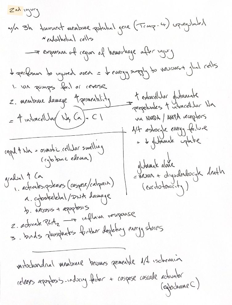
Secondary injury extends damage beyond the initial insult. The original “within 3 h” cue refers to TRPM4, correctly named transient receptor potential M4. In a rat spinal-contusion study, upregulation appeared at 3 h and later, especially in capillaries; no change was seen at 1.5 h. This is experimental evidence, not a universal timeline in dogs. TRPM4 carries monovalent ions, not calcium.
Reduced perfusion limits energy for neurons and glia. Ion pumps fail or reverse, membranes become more permeable, and intracellular sodium, chloride and calcium rise. Sodium accumulation draws water into cells: cytotoxic oedema.
Energy failure also reduces astrocytic glutamate uptake. Excess extracellular glutamate reinforces ion loading and can injure neurons and oligodendrocytes through excitotoxicity. NMDA and AMPA are reminders here; receptor contributions vary with cell type and experimental conditions.
The gradual calcium rise links three groups in the note: protease activity, PLA₂ and phosphate binding. Calpains are directly calcium-activated; caspases participate in regulated death cascades. Cytoskeletal damage, DNA injury, necrosis and apoptosis are related outcomes, not interchangeable terms. PLA₂ can support inflammatory lipid signalling, but its effects differ by enzyme form and model. The phosphate–energy line is a textbook summary: a cat study proposed calcium sequestration by inorganic phosphate, without directly proving ATP depletion from that process.
Ischaemic mitochondrial injury releases pro-death factors. Cytochrome c supports caspase-cascade activation, while apoptosis-inducing factor also has caspase-independent effects. These interacting mechanisms do not define a treatment window or a guaranteed clinical outcome.
References
Johnston & Tobias, 2nd ed., Ch 29; Gerzanich et al. 2009; Young & Koreh 1986; López-Vales et al. 2011; Susin et al. 1999.