
Primary and secondary CNS injury
Read the draft explanation
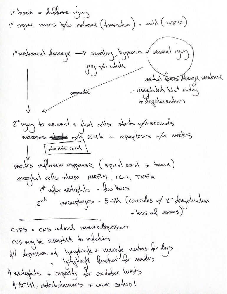
Primary injury is the initial mechanical damage. The original calls brain injury “diffuse” and places IVDD at the mild spinal end. Brain trauma commonly causes focal and diffuse changes; IVDD can also cause severe injury.
The map links mechanical damage with swelling, hypoxia and axonal injury in gray and/or white matter. Inertial forces can damage axonal membranes, allowing unregulated Na+ entry and depolarisation. Secondary injury then affects neurons and glia and can begin within seconds.
The book says most necrosis occurs in the first 24 hours, while apoptosis may continue for weeks. These are teaching timelines, not fixed clocks for every dog.
Inflammation begins rapidly and overlaps these cell-death processes. The cord-greater-than-brain comparison comes from matched experimental lesions in mice. MMP-9 upregulation is an early inflammatory event; it is not solely a microglial product. Microglia release IL-1 and TNF-α.
The note orders neutrophils first, within a few hours, then macrophages at 5–7 days, alongside secondary demyelination and axon loss. These are approximate book phases. Rat experiments report different timing; inflammation can also support repair.
CIDS means CNS injury-induced immunodepression. Injury can alter systemic immunity and increase susceptibility to infection. The days-long falls in lymphocyte and monocyte counts, and months-long reduction in lymphocyte function, come from cited human SCI/stroke work. They are not established canine schedules.
The note also recalls increased neutrophils and oxidative-burst capacity. In the cited rat experiment, the enhanced burst was transient; responses changed with time. The ACTH, catecholamine and urine-cortisol rise is an attributed stress-response reminder, not a fixed hormone pattern in every patient.
References
Johnston & Tobias, 2nd ed., Ch29; Schnell 1999; Crowe 1997 with corrigendum; Dusart 1994; Noble 2002; Cruse 1992; Riegger 2009; Gris 2008, abstracts.