
Cervical spondylomyelopathy - osseous and disc-associated patterns
Read the draft explanation
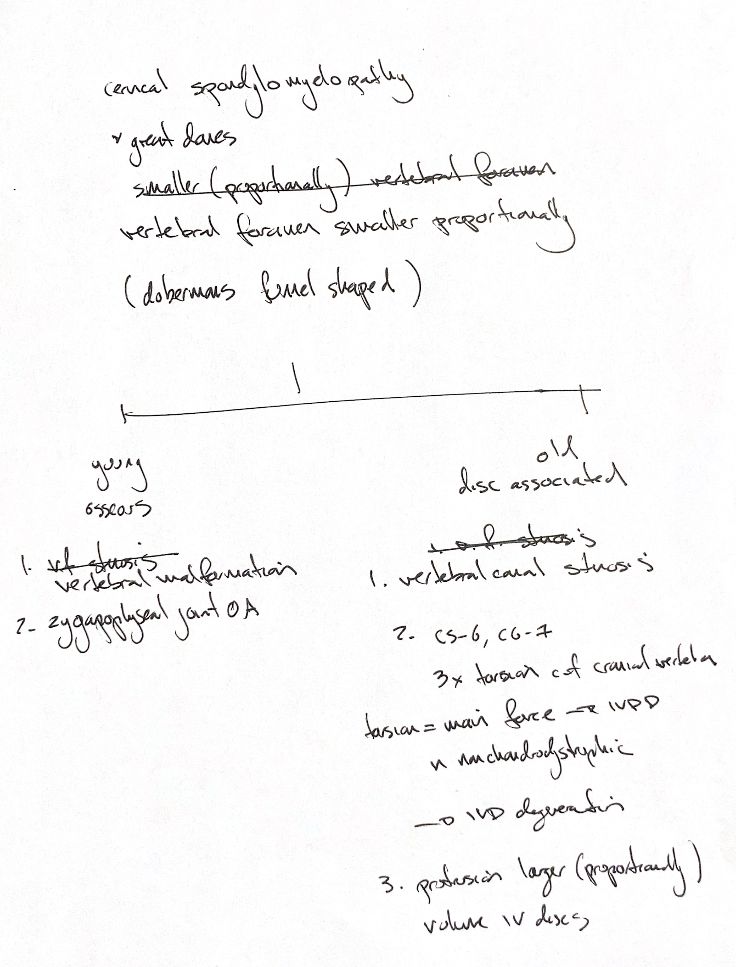
Cervical spondylomyelopathy, or wobbler syndrome, involves compression of the cervical spinal cord, nerve roots, or both. The vertebral foramen here means the opening that forms the vertebral canal, not the separate opening for a nerve root. Large and giant breeds can have proportionally less canal space. The source also recalls the funnel-shaped caudal cervical openings seen particularly in Dobermans. Less reserve space can contribute to compression; size alone does not establish the disease.
The young, osseous-associated side recalls vertebral malformation and osteoarthritis at the zygapophyseal joints. The older, disc-associated side recalls canal stenosis, the C5–C6 and C6–C7 levels, and protrusion of larger discs. These are typical patterns with overlap. Osseous and disc-associated compression can occur together.
The “3× torsion” cue is a rounded textbook reminder. Its cited experiment found about 2.6 times greater axial rotation in the caudal than cranial cervical segments of eight Foxhound cadavers. It is not a threefold disease risk in living dogs.
The original “torsion = main force” reminder needs a limit: the textbook cites a human lumbar cadaver paper here. Torsion is a proposed biomechanical contributor, not proof of a sole cause in nonchondrodystrophic dogs. The original larger-volume disc cue also needs care: the cited Doberman MRI study reported wider discs, rather than a direct volume measurement.
CSM is multifactorial. Age, breed, canal shape or an image alone cannot replace the complete clinical assessment.
References
Johnston & Tobias, 2nd ed., Ch31; Johnson et al. 2011; da Costa et al. 2006; Bonelli & da Costa 2023, indexed abstracts.