
Type I and II IVDD - mechanisms and historical limits
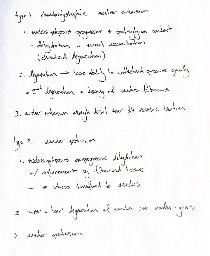
Read the draft explanation
The numbered chains describe the classic mechanisms behind nuclear extrusion and annular protrusion. The nucleus pulposus is the central pressure-bearing material; the annulus fibrosus is the surrounding fibrous ring.
In the Type I account, the nucleus progressively loses proteoglycans, dehydrates and accumulates mineral. Altered pressure distribution accompanies secondary annular degeneration and tearing. Nuclear material can then extrude through the annulus, often dorsally because the nucleus sits eccentrically. The original chondrodystrophic label is an association, not an exclusive breed rule. Both contusion and compression can contribute to injury.
The Type II chain describes progressive dehydration with replacement by “fibrinoid tissue”, the literal wording in the note and supplied textbook. Stress is transferred to the annulus; its wear-and-tear changes over months to years lead to protrusion. Those words preserve the historical explanation, not proof of a unique fibroid nuclear process in every nonchondrodystrophic dog.
Later histology challenges that strict cellular split. Hansen and colleagues studied 42 cadaver discs from 32 dogs without clinical signs of disc disease, examining grades I–III. Both breed groups showed chondroid changes, with no nuclear fibrocyte-like cells identified in those samples. That does not mean advanced scarring can never contain fibrocytes.
Keep the mechanical distinction: extrusion displaces internal disc material through the annulus; protrusion displaces the annulus beyond its normal boundary. Breed, tissue terminology and the degree of degeneration alone do not replace describing the actual herniation. The original two chains remain available beside these qualifications.
References
Johnston & Tobias, 2nd ed., Ch29; Hansen et al. 2017; Kranenburg et al. 2013; Fenn & Olby/CANSORT-SCI 2020.