
Cerebral blood flow, CPP and Cushing’s reflex
Read the draft explanation
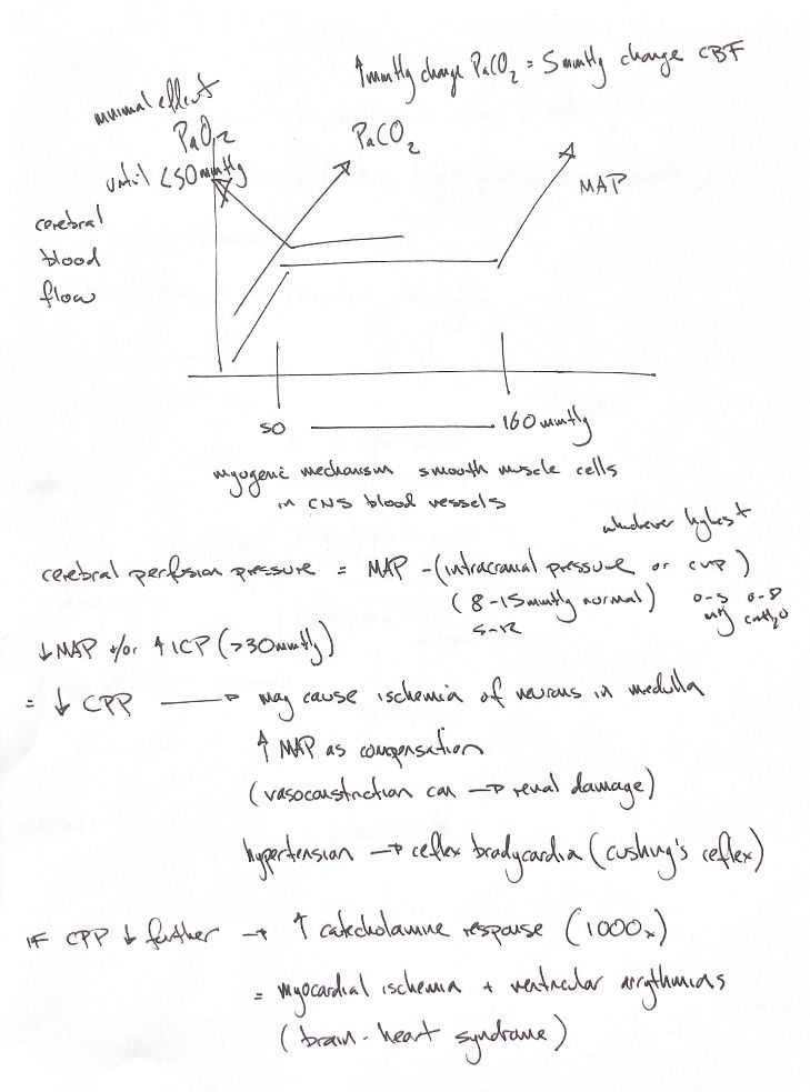
The three curves are schematic. The note and Ch29 place the MAP plateau at 50–160 mmHg; Ch36 shows 60–160 and describes roughly 60–180. These are different teaching ranges, not fixed limits for every patient. Myogenic vascular smooth muscle contributes to autoregulation, which brain injury can impair.
CO₂ strongly changes cerebral vascular tone. The original says 1 mmHg PaCO₂ changes CBF by “5 mmHg”. CBF is flow: Ch29 says about 5%, not 5 mmHg. The response depends on conditions. PaO₂ has less influence in its physiological range; the book shows a marked flow rise below 50 mmHg, not a safe oxygen threshold.
CPP=MAP−whichever is higher, ICP or CVP. Lower MAP or higher opposing pressure reduces this driving gradient. Flow also depends on vascular resistance. The note’s ICP 8–15 mmHg matches Ch29’s dog/cat estimate; Ch36 gives 5–12. Ch21 gives rounded CVP reference ranges of 0–5 mmHg or 0–8 cmH₂O.
The note pairs rising ICP with >30 mmHg. That passage concerns significant perfusion reduction in people; it is not a universal canine trigger or a pressure below which injury is excluded.
Severely reduced CPP may cause medullary ischaemia. A vasomotor response raises systemic pressure; intense vasoconstriction can damage other organs, including kidneys. Hypertension can then provoke baroreflex bradycardia: the classical Cushing response. Its absence does not exclude brain herniation.
With further compromise, a catecholamine surge can injure myocardium and provoke ventricular arrhythmias: brain–heart syndrome. The 1000× epinephrine report came after sudden ICP-induced brain death in 5 experimental dogs. This is not a typical clinical Cushing response or a fixed rise in every catecholamine.
References
Johnston & Tobias, 2nd ed., Ch21/29/36; Alberti et al. 1975; Artru et al. 1989; Grady & Blaumanis 1988; Her et al. 2022; Shivalkar et al. 1993.