
CT Hounsfield units and MRI signal cues
Read the draft explanation
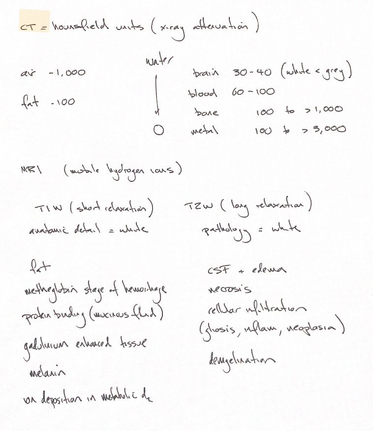
CT uses Hounsfield units, or HU, to describe x-ray attenuation relative to water at zero. Air is −1000 and fat is about −100. The brain is around 30–40, with white matter slightly below gray. The 60–100 blood cue concerns acute-to-subacute clotted blood. Bone and metal values vary. These numbers guide interpretation; they do not identify one substance on their own, and window settings change the displayed gray shades.
MRI describes relative signal intensity. T1W and T2W are image weightings, not a matching HU scale. Signal comes from mobile hydrogen protons in water and fat. T1 and T2 relaxation occur together; sequence timing changes which differences stand out. “Anatomic detail = white” and “pathology = white” are memory cues, not rules that every structure follows.
The T1-bright reminders are fat, the methemoglobin stage of hemorrhage, protein-rich or mucinous fluid, gadolinium-enhanced tissue, melanin and the original copper-deposition cue in metabolic disease. Appearance depends on composition, sequence and timing. Compare before and after contrast to separate enhancement from brightness already present. A bright T1 region alone cannot identify copper or diagnose melanoma.
On conventional T2W images, normal CSF and increased tissue water are often bright. The source lists edema, necrosis, cellular infiltration, gliosis, inflammation, neoplasia and demyelination. These appearances are variable and nonspecific. Not all pathology is bright, and some protein-rich or fibrotic tissues can be dark. FLAIR can suppress normal CSF signal.
Keep the numerical CT guide separate from the sequence-dependent MRI word lists, then interpret the complete study.
References
Johnston & Tobias, 2nd ed., Ch28/Table28.1; Michaelidou et al. 2024; Durant et al. 2025; canine indexed abstracts.