
Imaging - spatial resolution and MRI versus CT
Read the draft explanation
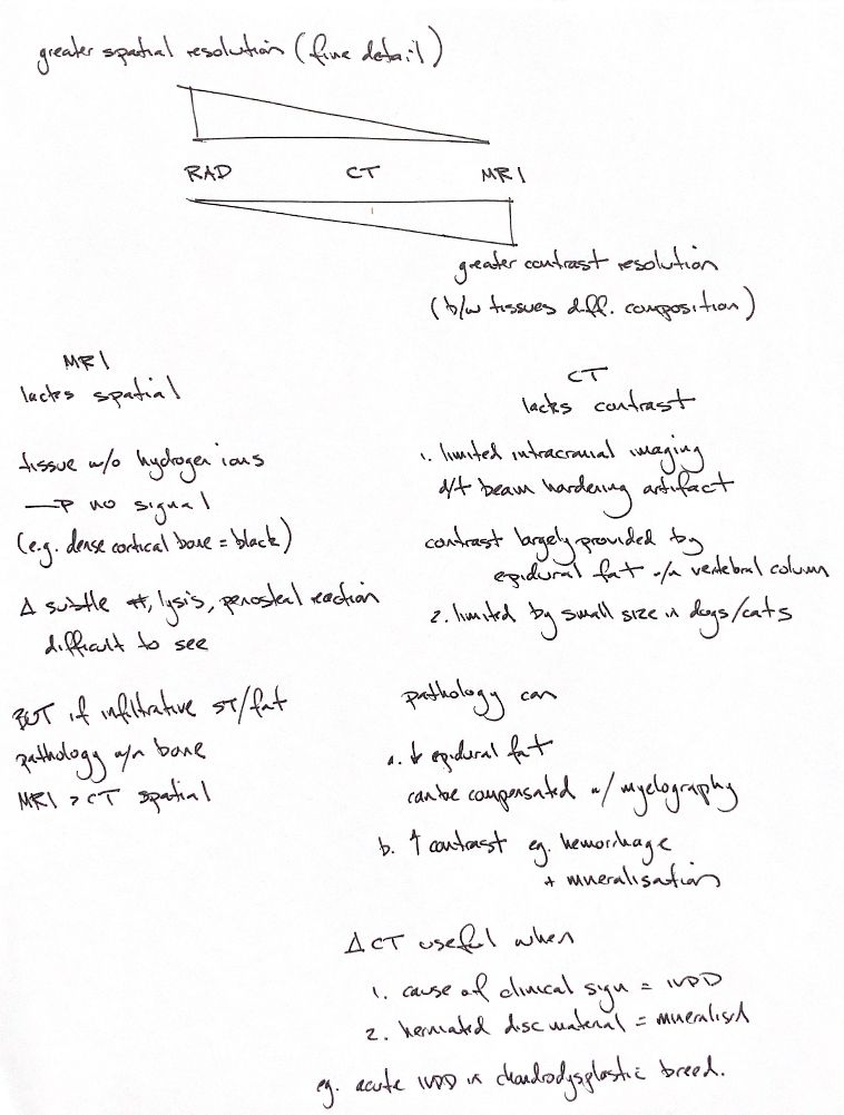
Spatial resolution means resolving fine detail; contrast resolution means distinguishing tissues of different composition. The opposing RAD–CT–MRI wedges are qualitative textbook reminders, not calibrated or universal rankings. Hardware and acquisition matter.
Conventional MRI usually shows less fine cortical detail than CT or radiography. Dense cortical bone has low detectable proton density and very short signal decay, so it appears dark on standard sequences. “Without hydrogen → no signal” is shorthand, not a literal claim that bone contains no hydrogen. Subtle fractures, lysis and periosteal reaction can be difficult to resolve.
Infiltrating soft tissue or replacement of normal marrow fat may nevertheless be conspicuous on MRI. The original “MRI > CT spatial” line needs that distinction: better lesion visibility through tissue contrast, rather than higher intrinsic spatial resolution.
CT has lower soft-tissue contrast than MRI. Beam hardening particularly affects the caudal cranial fossa. Within the small vertebral canal of dogs and cats, epidural fat helps outline structures. Fat effacement can obscure a lesion; CT myelography may help delineation. Acute blood or mineral may instead increase attenuation and lesion contrast.
The final CT cue assumes the signs come from disc herniation and the extruded material is mineralised, illustrated by acute disease in a chondrodystrophic dog. That combination can favour plain CT, but does not guarantee a diagnostic study. Uncertain assumptions, nonmineralised lesions or cord disease may need MRI for better characterisation.
References
Johnston & Tobias, 2nd ed., Ch28; Cooper et al.2014; Cicci et al.2025; Gielen et al.2013 and Zarelli et al.2014, indexed abstracts.