
Spinal reflex segments and reliability
Read the draft explanation
A spinal reflex tests its sensory and motor arc. These are spinal cord segments, not vertebral levels.
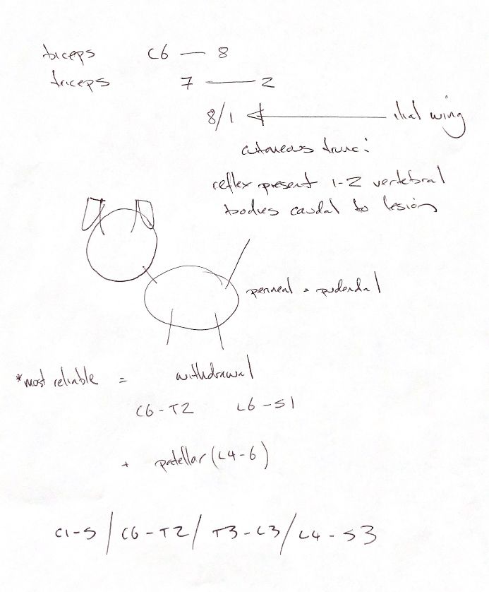
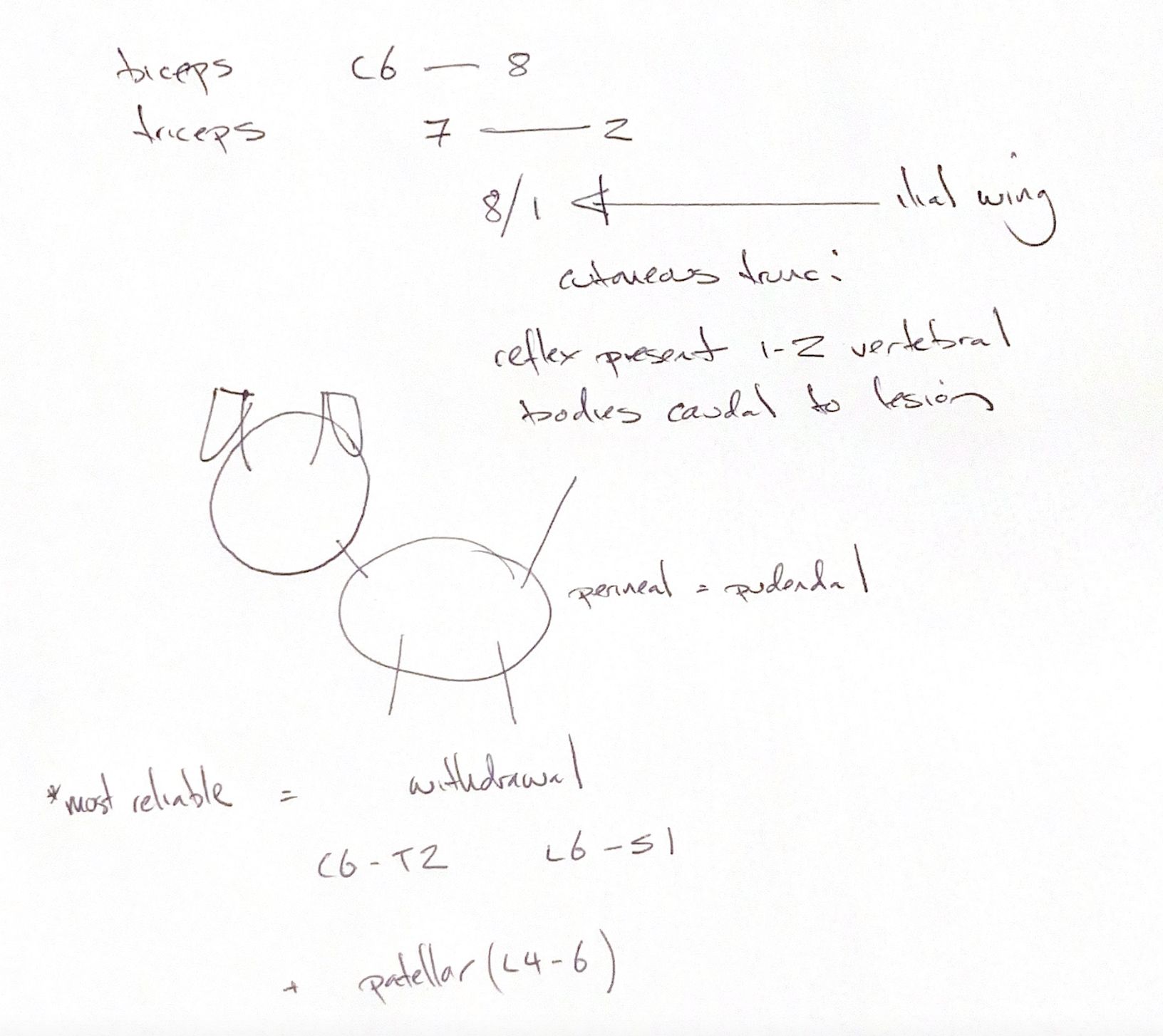
The original and supplied textbook map biceps to C6–C8 and triceps to C7–T2. Their most reliable routine reflexes are withdrawal and patellar: thoracic withdrawal C6–T2, sciatic-mediated pelvic withdrawal L6–S1, and patellar L4–L6. Biceps and triceps responses can be difficult to elicit in normal animals. Reliability is not a guarantee in every patient.
Published maps are not identical. The WSAVA table gives triceps C7–T1 and broader pelvic withdrawal L4–S2, alongside narrower sciatic contributions. Match the segment range to the component being assessed rather than treating each range as interchangeable.
For cutaneous trunci, C8/T1 is the textbook’s cervicothoracic motor relay. The iliac wing is an approximate caudal skin landmark. They are different locations. Other canine lateral-thoracic nerve descriptions include T2; they do not put these cord segments at the pelvis.
The textbook says the reflex can remain present 1–2 vertebral bodies caudal to a lesion, depending on its location along the thorax. This is a localisation clue, not a fixed ruler. Some normal dogs and cats do not show the reflex.
The perineal reflex uses the pudendal nerve, with sacral and caudal cord contributions. The four broad localisation regions in the note are C1–C5, C6–T2, T3–L3 and L4–S3.
Interpret reflexes with muscle tone, muscle mass and the rest of the examination. Limb withdrawal is a spinal reflex; it does not by itself demonstrate conscious pain perception.
References
Johnston & Tobias, 2nd ed., Ch26; University of Illinois veterinary neurological examination; Berendt, WSAVA2017; MSD Veterinary Manual; Sharp et al., 1991.