
Horner syndrome, strabismus, nystagmus and PLR
Read the draft explanation
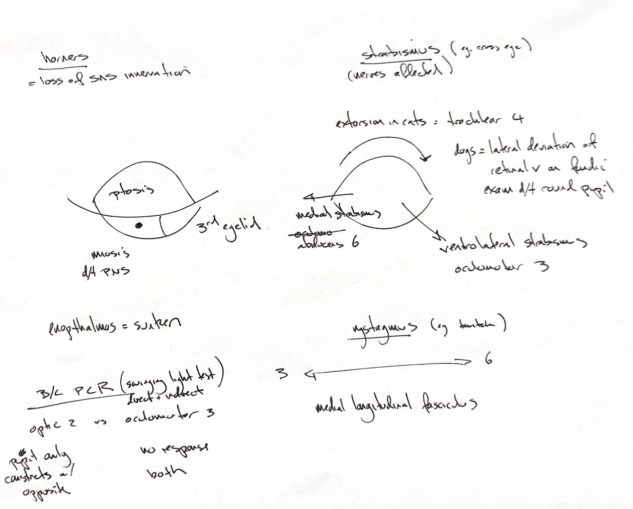
Horner syndrome reflects loss of sympathetic supply to the eye: ptosis, a raised third eyelid, miosis and enophthalmos. The small pupil reflects loss of dilator drive with parasympathetic constriction relatively unopposed. Enophthalmos means the globe sits further back in the orbit.
For the original strabismus cues, III is associated with ventrolateral deviation, VI with medial deviation, and IV with globe extorsion. Torsion is easier to see in the cat’s pupil; in dogs, the round pupil makes the retinal-vein orientation on fundic examination useful. These are nerve-function associations, not proof of one isolated lesion. The two original eye sketches have no declared patient-side projection and are kept separate.
The note’s III↔VI and medial longitudinal fasciculus cue belongs within vestibular eye-movement coordination. CN VIII provides vestibular input; the brainstem links it with ocular motor nuclei. Physiological nystagmus with head movement and pathological nystagmus are not interchangeable diagnoses.
The bottom comparison concerns the affected pupil. If it fails to constrict when its own eye is lit but constricts when the opposite eye is lit, the textbook considers ocular, retinal or optic afferent dysfunction, provided the opposite eye is normal. If it remains dilated with either eye lit, parasympathetic III/nuclear dysfunction or iris-muscle disease must be considered. Observe direct and consensual responses in both eyes and interpret the entire ocular and neurological examination; this shorthand alone does not identify a single nerve lesion.
References
Johnston & Tobias, 2nd ed., Ch26, Table26.1 and ocular/vestibular examination sections; University of Guelph cranial nerve teaching notes; University of Minnesota autonomic teaching table.