
Cranial nerve exits and the alar-foramen correction
Read the draft explanation
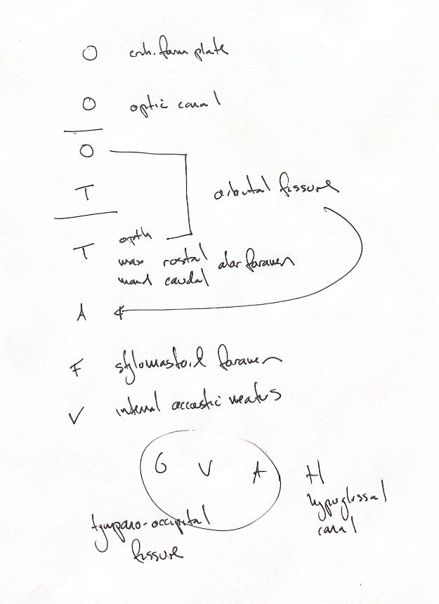
The letters follow cranial nerves I to XII. Olfactory fibres pass through the cribriform plate, and the optic nerve uses the optic canal. The orbital fissure is shared by the oculomotor, trochlear and abducent nerves, plus the ophthalmic branch of the trigeminal nerve. My long curved pointer links that shared exit back to the abducent row; it is not a drawn nerve course.
The trigeminal branches need one correction. In the dog, the maxillary nerve passes through the round foramen into the alar canal, then leaves by the rostral alar foramen. The mandibular nerve uses the oval foramen. My original mandibular “caudal alar” cue is corrected here, with the original kept alongside the set.
The facial nerve passes through the internal acoustic meatus and facial canal before reaching the skull surface at the stylomastoid foramen. The vestibulocochlear nerve uses the internal acoustic meatus. These are different levels along the facial nerve's route.
The circled G, V and A stand for glossopharyngeal, vagus and accessory. They pass through the jugular foramen and tympano-occipital fissure. The hypoglossal nerve has its own hypoglossal canal. These are canine exit relationships; the source does not show a full skull or nerve atlas.
References
Johnston & Tobias, 2nd ed., Ch122 (facial route); Fletcher & Clarkson, veterinary gross anatomy, pp44–46; NC State cranial-nerve lab; Santifort et al.2023, selected canine anatomical text.