
Cranial nerves: modality and brain regions
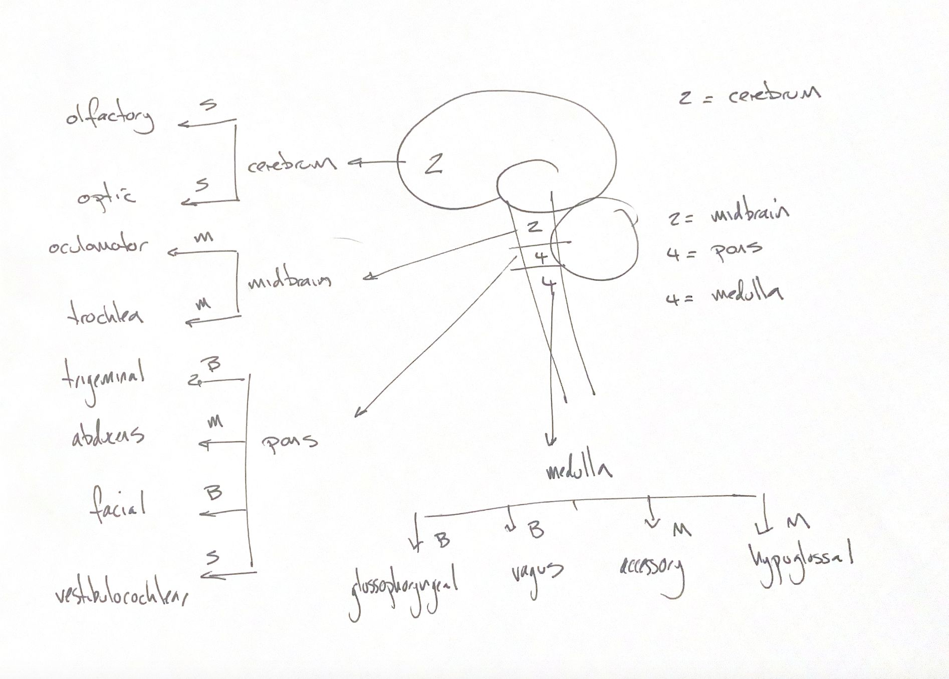
Read the draft explanation
S means sensory, M means motor, and B means both. These are simple functional labels. They do not list every fibre type carried by a nerve.
The sensory group is I olfactory, II optic and VIII vestibulocochlear. The motor group is III oculomotor, IV trochlear, VI abducens, XI accessory and XII hypoglossal. V trigeminal, VII facial, IX glossopharyngeal and X vagus have both sensory and motor functions. Motor function can include parasympathetic output, as it does in III.
My 2–2–4–4 cue groups two with cerebrum, two with midbrain, four with pons and four with medulla. It is a recall map, not an exact map of canine attachments or all cranial nerve nuclei.
I carries axons from olfactory receptor neurons in nasal epithelium to the olfactory bulb. II carries retinal ganglion-cell axons and is associated with the diencephalon. Calling both simply “cerebrum” misses that distinction.
III and IV are associated with midbrain. V attaches at pons, but its nuclei extend into other regions. Canine anatomy places the emergence of VI, VII and VIII at the rostral medulla. They should not all be assigned to pons just because the source groups them there.
IX, X and XII have medullary associations. XI also has a cervical spinal component. The traditionally described bulbar component joins the vagus. A nerve’s attachment, its nuclei and the cell bodies that supply its sensory axons are related, but different, questions.
References
Johnston & Tobias, 2nd ed., Ch26, Table26.1; University of Minnesota veterinary neuroanatomy and canine brain atlas; University of Guelph cranial nerve teaching notes.