
Meninges, CSF and hydrocephalus
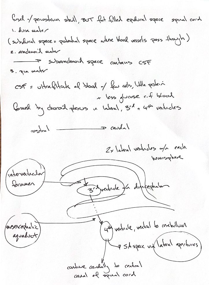
Read the draft explanation
From outside inward, the meninges are dura mater, arachnoid mater and pia mater. Cranial dura is fused to the skull periosteum. In the vertebral canal, a fat-filled epidural space lies outside the dura. CSF occupies the subarachnoid space between arachnoid and pia.
My note and the textbook call the subdural region a “potential space” through which vessels pass. Microscopic reviews describe no normally open compartment at the dura–arachnoid junction. A collection can split the dural border-cell layer. The vessel wording does not mean that vessels sit in a normal open space.
Normal CSF has few cells, little protein and less glucose than blood. The original “ultrafiltrate” shorthand needs a qualifier: the choroid plexus epithelium actively secretes and regulates CSF. It is not simply a passive blood filtrate. The choroid plexuses in the lateral, third and fourth ventricles are the main source; this is not an exclusive-source claim.
The drawing follows the usual bulk-flow route. There are two lateral ventricles, one in each cerebral hemisphere. CSF passes through the interventricular foramina to the third ventricle within the diencephalon, then through the mesencephalic aqueduct to the fourth ventricle, ventral to the cerebellum. It can enter the subarachnoid space through the lateral apertures or continue caudally into the spinal central canal.
The interventricular foramina, mesencephalic aqueduct and lateral apertures are normally narrow. Disease can obstruct these sites and lead to hydrocephalus. My circles mark locations, not diagnosed blockages.
References
Johnston & Tobias, 2nd ed., Ch29; Haines et al.1993 and Pollay et al.1985, indexed abstracts; Vernau et al.2008; MacAulay et al.2022, selected text.