
Brain regions and cerebral connections
Read the draft explanation
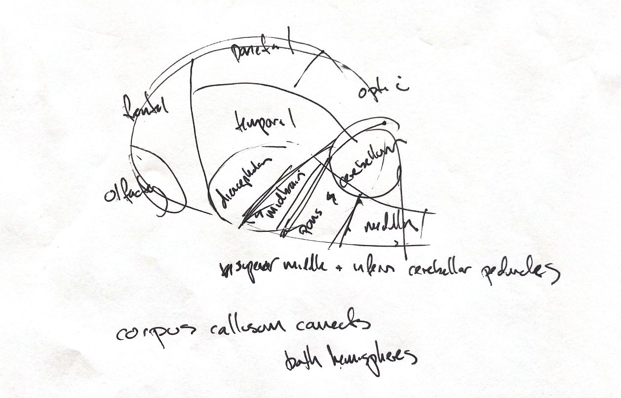
The first sketch is a simple map of the cerebral lobes and nearby brain regions. It keeps the frontal, parietal, temporal and olfactory labels, with the diencephalon, midbrain, pons, medulla and cerebellum. My posterior cerebral label says “optic”. The standard lobe name used here is occipital. These coarse borders are memory cues, not a full anatomical atlas or a map of each region's function.
Three cerebellar peduncles connect the cerebellum with the brainstem. My superior, middle and inferior terms correspond to rostral, middle and caudal in veterinary anatomy. The rostral peduncle links with the midbrain, the middle with the pons, and the caudal with the medulla. The drawn pointers mark connections. They do not mean that every axon carries information in the same direction.
The corpus callosum connects the two cerebral hemispheres. It is a white-matter commissure: a bundle of axons crossing between them. This is a separate connection from the cerebellar peduncles.
References
Johnston & Tobias, 2nd ed., Ch29/35; University of Minnesota veterinary neuroanatomy, Cerebellar Peduncles and Telencephalon.