
Pericardial volume and cardiac tamponade
Read the draft explanation
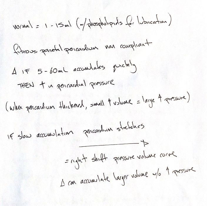
My note and Chapter 107 give 1–15 mL of normal canine pericardial fluid. Chapter 106 instead gives 0.5–1 mL. The book also gives 5–60 mL as an acute accumulation cue for rising pressure. These are textbook estimates, not validated reference intervals or universal thresholds. The rate of accumulation and pericardial stiffness matter.
Rapid accumulation meets a fairly noncompliant fibrous sac. A thickened pericardium can develop a large pressure rise with little added volume. Slow accumulation allows stretch and shifts the pressure–volume relationship rightward. More fluid can then accumulate before pressure rises sharply; that reserve is still limited.
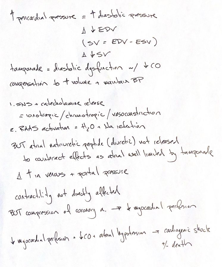
As pericardial pressure rises, pressure measured inside the heart can rise while its effective distending pressure falls. Filling and EDV fall, reducing stroke volume and cardiac output. SV=EDV−ESV remains the volume relationship. Tamponade restricts diastolic filling; it does not require primary loss of myocardial contractility.
Sympathetic activity and catecholamines increase heart rate, contractile drive and vascular tone. RAAS retains sodium and water. These responses try to maintain circulating volume and blood pressure. Systemic venous and portal pressures can rise.
The ANP shorthand needs care. Constrained atrial stretch can limit the expected natriuretic and diuretic response despite high atrial pressure. Canine studies found stable or non-elevated plasma ANP during tamponade, not an absence of the peptide.
My final cue links coronary compression with reduced myocardial perfusion. Coronary flow also depends on arterial pressure and neurohumoral responses. Poor perfusion, low output and hypotension can lead to shock and death. The source calls this cardiogenic shock; the initiating problem here is external restriction of filling.
References
Johnston & Tobias, 2nd ed., Ch106/107; Santamore1990; Zioris1989; Klopfenstein1990; Stokhof1994; Nakata1978; Bernath1987, indexed abstracts.