
Heart-failure compensation and the renin–angiotensin–aldosterone system
Read the draft explanation
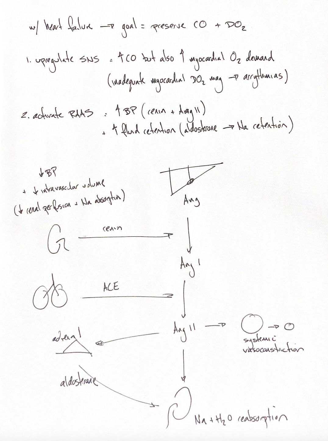
When cardiac output falls, the body tries to preserve blood flow and oxygen delivery. The sympathetic nervous system can raise cardiac output, but it also raises myocardial oxygen demand. If oxygen delivery to the myocardium is inadequate, arrhythmias may follow.
The renin–angiotensin–aldosterone system, or RAAS, supports blood pressure and retains sodium and water. Reduced renal perfusion and reduced NaCl signalling at the macula densa can stimulate renin release. Sympathetic stimulation can also activate this system. The macula densa signal is separate from the increased renal sodium reabsorption shown at the end of the pathway.
The kidney releases renin, an enzyme that cleaves liver-derived angiotensinogen to angiotensin I. Angiotensin-converting enzyme, or ACE, then forms angiotensin II. The lung cue marks an important site of ACE activity; ACE also occurs elsewhere.
Angiotensin II constricts systemic vessels. It also promotes renal sodium retention directly and stimulates aldosterone release from the adrenal cortex. Aldosterone further increases renal sodium and water reabsorption. Both kidney branches in the drawing matter.
Low intravascular volume can trigger RAAS, but heart failure does not always mean low total fluid volume. Reduced effective arterial filling can coexist with sodium retention, fluid expansion and congestion. Hormonal responses vary with the stage and model of heart failure. Compensation can support circulation at first, while persistent vasoconstriction and retention can add to the burden on the heart.
References
Johnston & Tobias, 2nd ed.; Bugge et al. 1988; Hall et al. 1980; Watkins et al. 1976.