
ECG in relation to pressure volume loop
Read the draft explanation
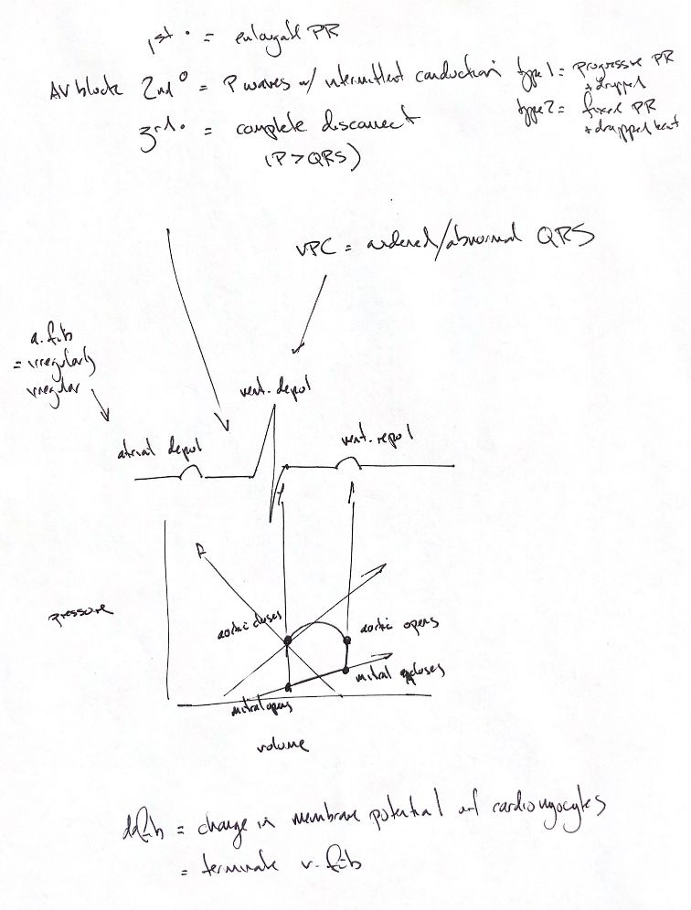
The ECG records electrical activity. The P wave represents atrial depolarisation, QRS ventricular depolarisation and T ventricular repolarisation. Electrical activation precedes the associated mechanical response; an ECG alone does not establish effective circulation.
In the idealised left-ventricular cycle, mitral closure begins isovolumetric contraction. Aortic opening permits ejection. Aortic closure begins isovolumetric relaxation, and mitral opening permits filling. The pressure-volume loop plots pressure against volume, so its horizontal axis is not time. The original cross-plot leaders are preserved in the source companion, but are not used as calibrated ECG-to-valve timing.
The upper notes distinguish AV block. First degree prolongs the PR interval. Second degree intermittently leaves a P wave without a conducted QRS: the typical Mobitz I pattern progressively prolongs PR before a dropped complex, while Mobitz II has unchanged conducted PR intervals. Third degree means no atrial impulses conduct to the ventricles: atrial and ventricular rhythms are independent. The note’s P > QRS reminder refers to rate or count, not waveform amplitude; that comparison alone does not establish complete block.
Atrial fibrillation commonly gives an irregularly irregular ventricular rhythm with absent discrete P waves. Irregularity alone is not diagnostic. A ventricular premature complex arises prematurely from the ventricles and usually has a wide, abnormal QRS; width alone is insufficient. The single drawn beat is a schematic, not an example of both arrhythmias.
Defibrillation changes myocardial membrane potentials and can interrupt ventricular fibrillation. It is not uniform depolarisation of every cell, and terminating fibrillation does not itself prove effective circulation.
References
Johnston & Tobias, 2nd ed.; Merck Veterinary Manual, Kittleson, updated 2025; Witkowski et al. 1990; Li et al. 1993; Efimov et al. 1997.