
Muscles of inspiration and expiration
Read the draft explanation
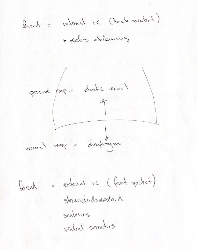
The diaphragm provides the main inspiratory movement at rest. It contracts and moves caudally, increasing thoracic volume. When it relaxes, elastic recoil helps the lungs return toward their earlier volume. The upward and downward arrows show movement, not the direction of airflow.
My passive-expiration cue describes the recoil mechanism. It does not mean all quiet expiration in dogs is passive. In a study of eight conscious dogs, abdominal muscles were active during quiet expiration, especially the transversus abdominis. The pattern changed with posture.
For forced expiration, my note pairs internal intercostals with rectus abdominis. “Back pocket” is the internal-intercostal memory cue. That broad pattern does not describe every individual muscle region. The parasternal, or interchondral, part of the internal intercostals helps inspiration. Canine intercostal effects vary along the rib cage and with neural recruitment.
For increased inspiratory demand, my source and the book list external intercostals, sternocleidomastoid, scalenus and ventral serratus. “Front pocket” is the external-intercostal cue. These are the source’s accessory-muscle terms, rather than a universal canine recruitment pattern.
A canine study tested scalene and sternomastoid activity in 18 anaesthetised dogs and three awake dogs. It found little or no respiratory activation under the tested conditions. That result limits a blanket neck-muscle claim; it does not describe every species or situation. The study measured sternomastoid, while the source’s list uses sternocleidomastoid.
References
Johnston & Tobias, 2nd ed., Ch103; De Troyer et al. 1989, 1994 and 1999; De Troyer & Farkas 1993.