
Aortic arch branches and thoracic venous return
Read the draft explanation
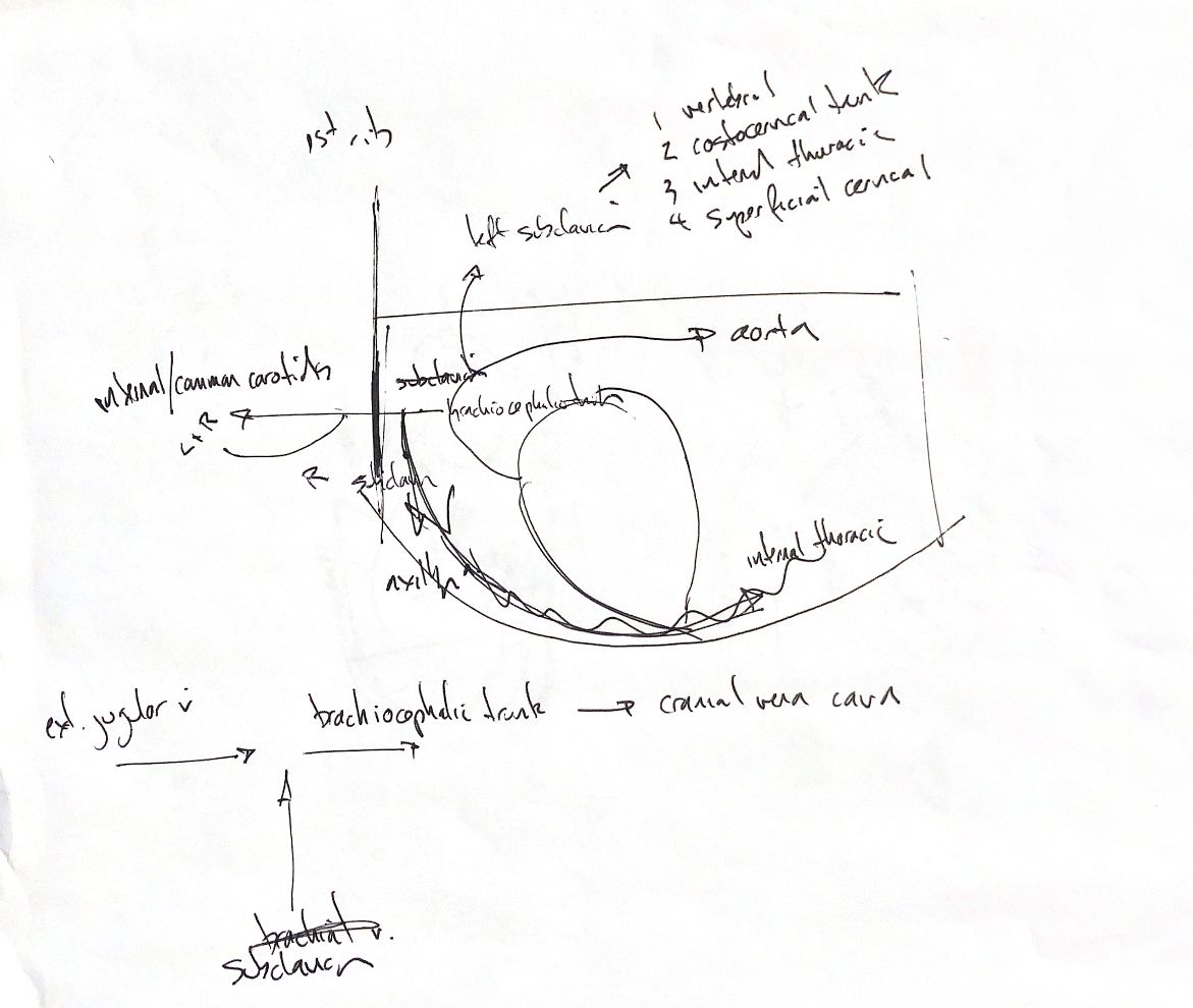
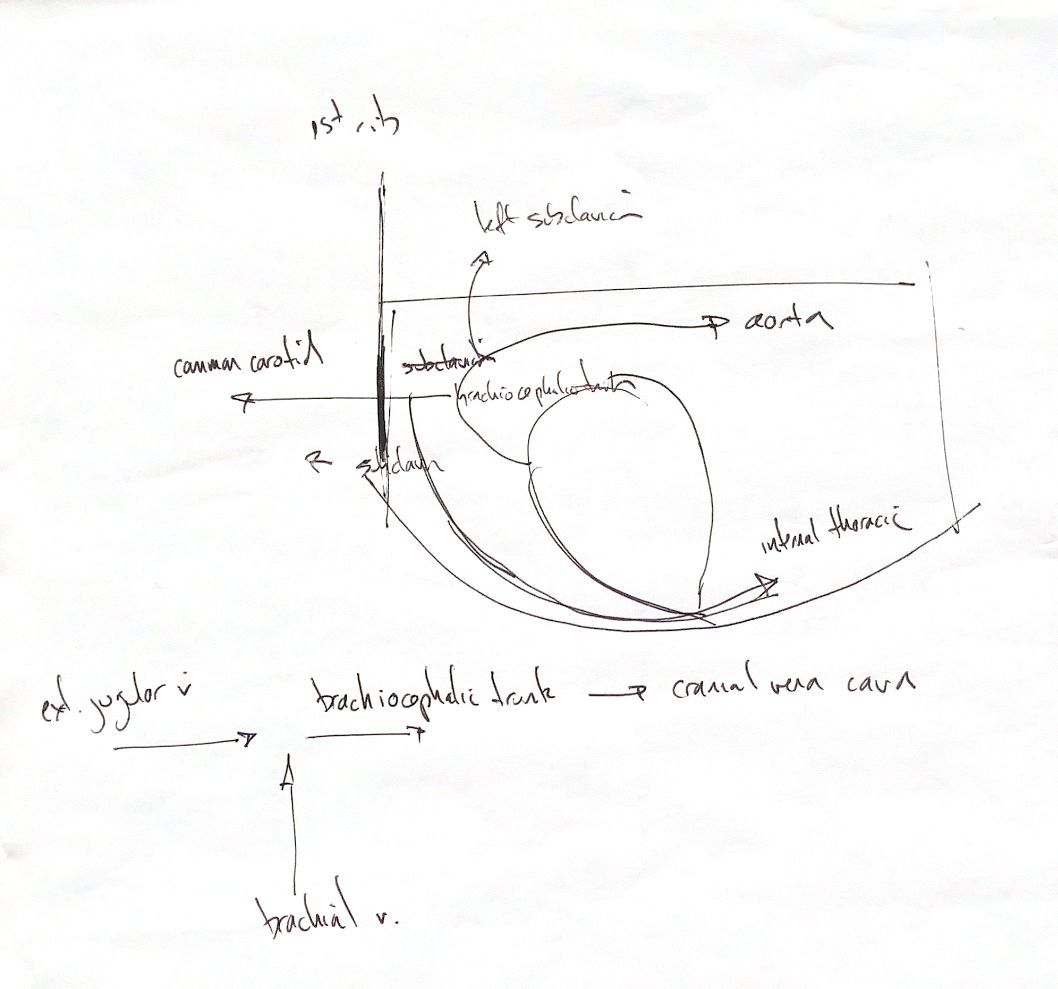
My upper drawing is a lateral thoracic sketch. Cranial is to the left of the page; caudal is to the right. The vessel names refer to the patient’s sides.
In the usual dog and cat pattern, the aortic arch gives off the brachiocephalic trunk first, then the left subclavian artery. The trunk supplies the left and right common carotid arteries and the right subclavian artery. The two subclavian arteries therefore have different origins. My original “external/common carotids” wording needs care: the branches from this trunk are common carotids, not external carotid origins.
My first drawing also lists four subclavian branches: vertebral, costocervical trunk, internal thoracic and superficial cervical. The numbering is a study list, rather than a fixed order in every animal. These branches arise before the subclavian becomes the axillary artery at the first rib. The internal thoracic artery runs caudally beside the sternum.
The lower map is venous return. The external jugular and subclavian veins join on each side to form a brachiocephalic vein. The two brachiocephalic veins then unite as the cranial vena cava.
There is a wording conflict in the sources. One drawing crosses out “brachial vein” and replaces it with “subclavian”; the other still says brachial. The textbook paragraph also uses brachial veins and brachiocephalic trunks. The teaching map uses the verified external-jugular/subclavian junction and the name brachiocephalic vein. That vein is separate from the arterial brachiocephalic trunk. Both originals remain available unchanged.
References
Johnston & Tobias, 2nd ed., Ch90,104–106; University of Minnesota dog/cat dissection guide; NC State cardiovascular anatomy.