
Lung lobes, rib neurovascular bundle and pulmonary vessel positions
Read the draft explanation
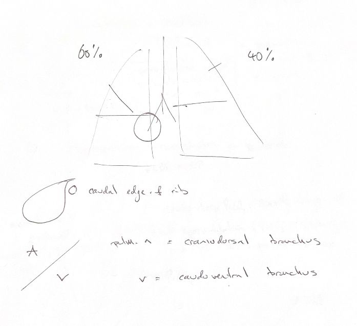
In dogs and cats, the right lung has cranial, middle, caudal and accessory lobes. The left lung has cranial and caudal lobes. Its cranial lobe has cranial and caudal parts, which share a common lobar bronchus. These are two parts of one lobe, rather than a separate left middle lobe.
The accessory lobe belongs to the right lung. It lies medially and passes dorsal to the caudal vena cava. A lobe’s bronchial supply helps define its identity, so an external fissure does not always mean a separate lobe.
In the dog, the right lung is larger. My 60/40 pair is a rounded size cue. The book gives 42% for the left lung’s share of lung mass. Measured volume proportions vary with the population and method, so 60/40 is not a fixed measured ratio.
The main intercostal nerve and vessels run along the caudal edge of each rib. Within an intercostal space, that means they lie just caudal to the cranial rib.
The pulmonary artery cue is craniodorsal to the bronchus; the pulmonary vein cue is caudoventral. These describe the lobar relationship. Central pulmonary vessels take more complex courses around the main bronchi, so the cue does not replace the hilar anatomy.
References
Johnston & Tobias, 2nd ed., Ch103–104; University of Minnesota, Carnivore Anatomy Lab11; Hsia et al. 1993/1994; Drees et al. 2011.