
Vessel branching, veins and vessel walls
Read the draft explanation
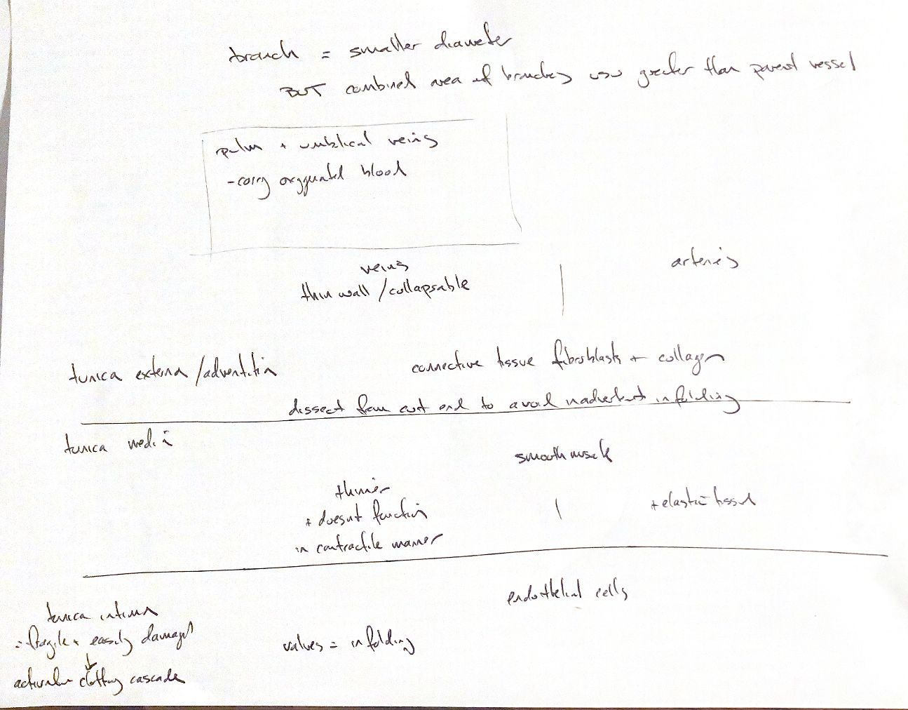
A branch has a smaller diameter than its parent artery. When one artery divides into two, the book says their combined area is generally greater than the parent's. This means cross-sectional area through the lumens, not the outer wall's surface area. It is a general comparison, not a fixed rule for every branch.
Veins carry blood toward the heart. Oxygen content does not define them. Pulmonary veins carry oxygenated blood from the lungs, and the fetal umbilical vein carries oxygenated blood from the placenta to the fetus. This differs from the usual systemic venous return.
Veins generally have thinner, more collapsible walls than comparable arteries. Both have three main layers. The outer tunica externa, or adventitia, contains connective tissue, fibroblasts and collagen. The middle tunica media contains smooth muscle; arterial media also contains elastic tissue. Venous media is thinner. The inner tunica intima has an endothelial lining, with its supporting basement membrane and connective tissue. The balance varies with vessel type and size.
My note and the book describe venous media as generally noncontractile. That is a simplification: venous smooth muscle can contract. Isolated canine saphenous veins contract in response to norepinephrine and serotonin. A thin wall does not mean inactive muscle.
The surgical cues are to keep adventitia from folding into the lumen during reconstruction and to protect the fragile intima. Intimal injury can activate the clotting cascade and cause thrombosis. This is a risk, not an inevitable result of every injury. Venous valves are infoldings of the intima. Where present, they help prevent backflow and pooling; not every vein has valves.
References
Johnston & Tobias, 2nd ed., Ch108; Curro et al.1978; University of Leeds and Columbia histology guides; University of Minnesota veterinary embryology.