
TECA: major vessels and nerves
Read the draft explanation
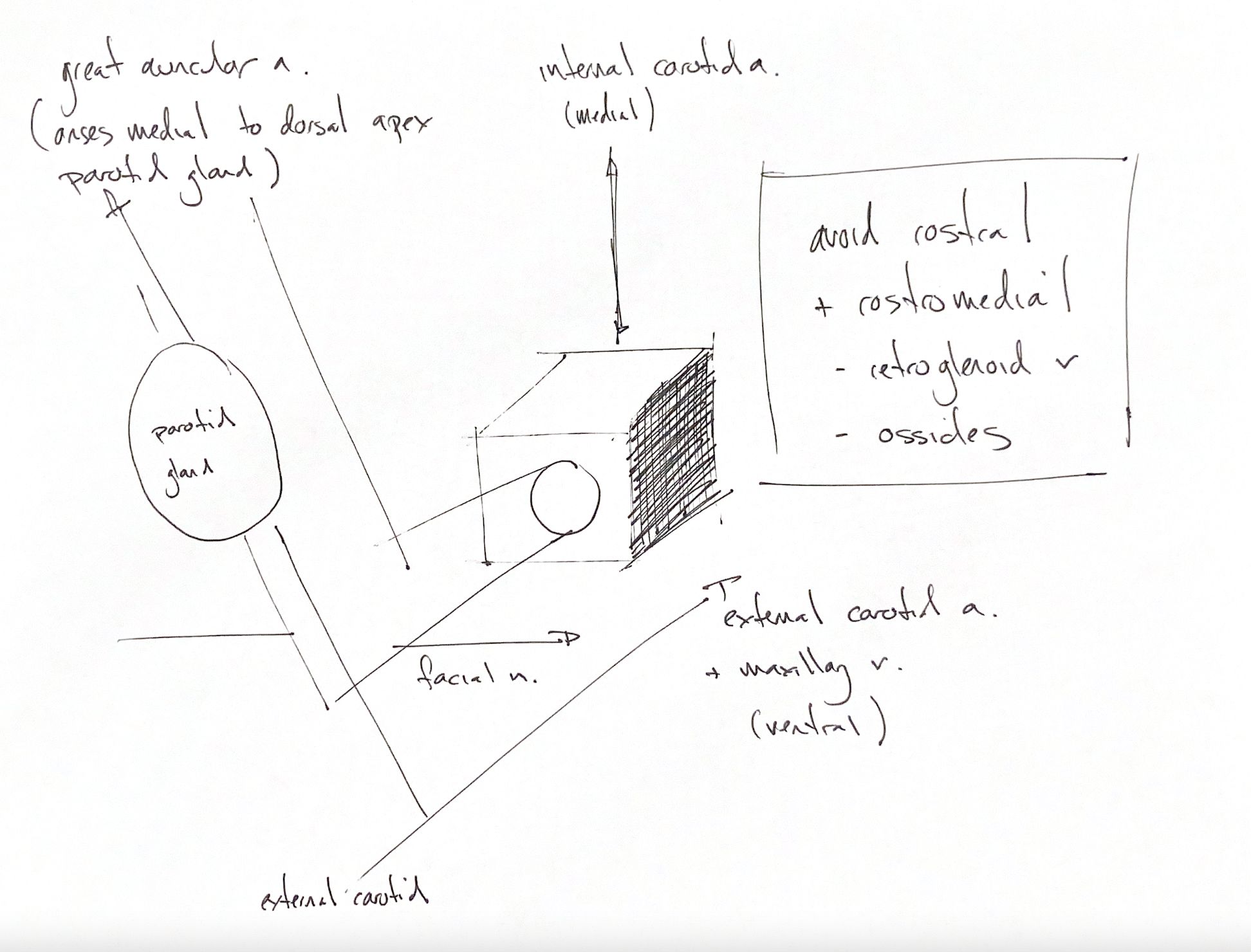
During a total ear canal ablation, the parotid gland is a useful landmark. The great auricular artery branches from the external carotid artery. It arises medial to the dorsal apex of the parotid gland, which overlies the vertical ear canal.
The facial nerve is cranial nerve VII. In the dog, it leaves the skull through the stylomastoid foramen, caudodorsal to the bony ear canal. It then crosses the ventral aspect of the horizontal canal. Its exit and its later course are separate landmarks.
The internal carotid artery lies immediately medial to the tympanic bulla. The external carotid artery and maxillary vein lie immediately ventral to it. Excessive debridement can collapse the medial bulla wall, and infection or neoplasia can erode it. That puts the medial artery at risk.
My boxed “avoid rostral + rostromedial” cue groups the retroglenoid vein and ossicles. Their positions are distinct. The retroglenoid vein is immediately rostral to the osseous ear canal. Straying rostrally during dissection can damage it. The auditory ossicles lie dorsally within the tympanic cavity. The textbook describes sparing visible ossicles in minimally affected ears; severe disease may hide them.
The drawing is a regional memory map. Its simple block and unlabelled circle are not a scale dissection or a map of the facial nerve's exit.
References
Johnston & Tobias, 2nd ed., Ch122, Fig122.2.