
Tympanic membrane: structure, self-cleaning and repair
Read the draft explanation
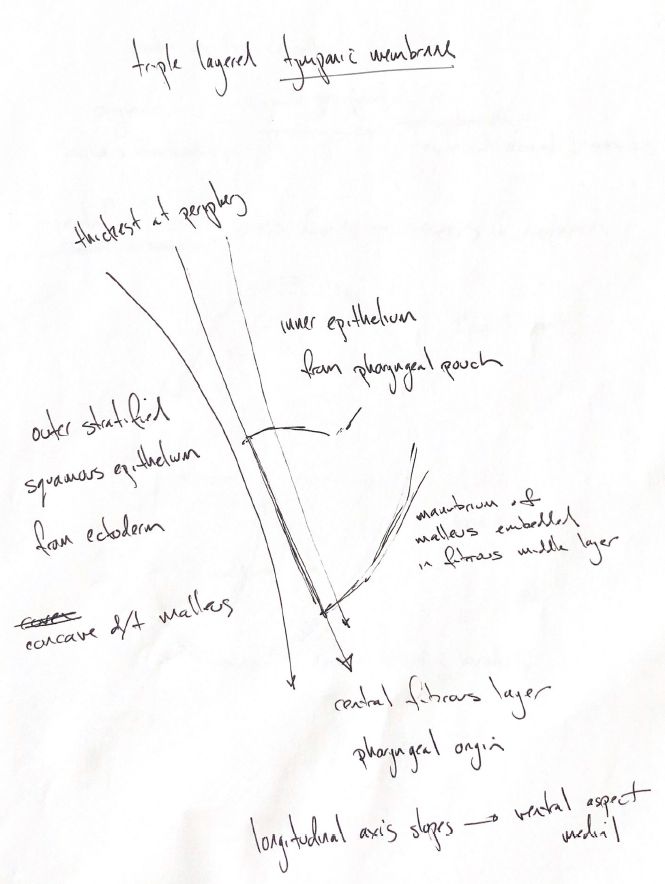
The tympanic membrane has three layers. The outer stratified squamous epithelium comes from ectoderm. The inner epithelium comes from the pharyngeal pouch. Between them is the central fibrous layer, described in the book as having pharyngeal origin.
The manubrium, or handle, of the malleus lies in the fibrous middle layer. Its attachment makes the outer surface concave. The membrane also slopes, with its ventral aspect lying more medially.
My “thickest at the periphery” cue recalls that thickness varies across the membrane. The book's cited measurements were made in fresh cat eardrums. They support peripheral thickening, rather than one fixed thickness for every dog.
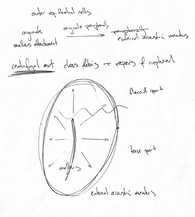
The smaller dorsal part is the pars flaccida. The larger ventral part is the pars tensa. The outer epithelial cells move from the malleus attachment toward the external acoustic meatus in the book's description. This carries surface debris outward.
In this note, centrifugal means outward. Paths can be straight or curved. Ink markers in healthy dogs confirmed outward movement. That experiment did not trace every cell's origin or test healing of ruptured membranes.
Epithelial migration also contributes to repair after rupture. It does not guarantee that every perforation closes normally or within a fixed time. The arrows show a movement pattern, not a treatment or a healing deadline.
References
Johnston & Tobias, 2nd ed., Ch123; Tabacca2011; Kuypers2005; Truy1995.