
Inner ear, balance and hearing
Read the draft explanation
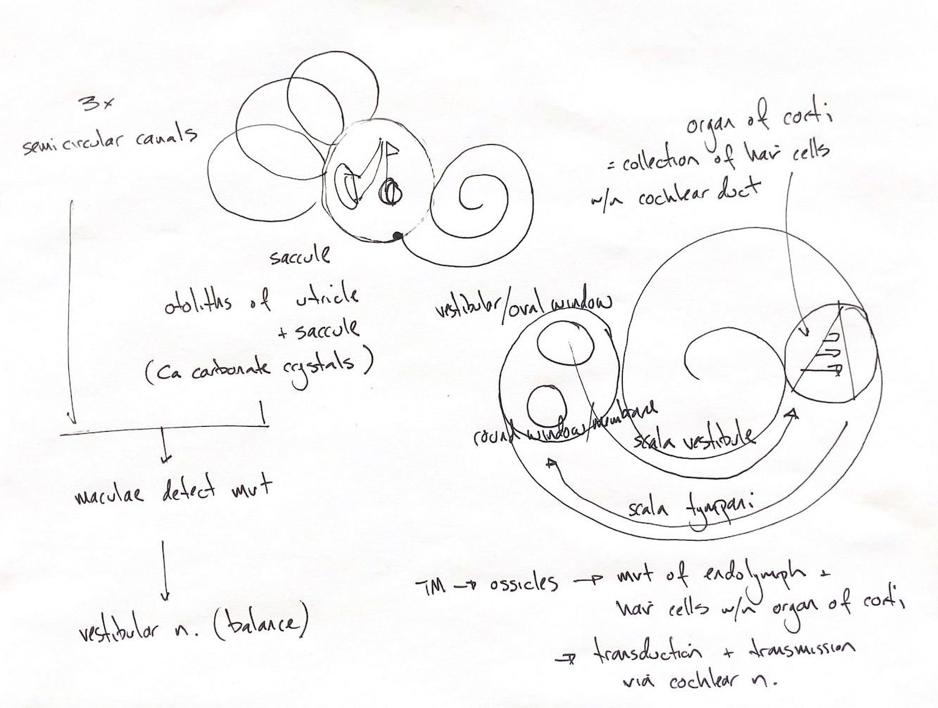
My overview links three semicircular canals, the vestibular region and the spiral cochlea. It is a memory sketch, not an exact view or scale. The vestibule contains both the utricle and saccule. The small internal marks are a memory cue, not named anatomy.
Balance uses two kinds of movement sensor. The utricle and saccule contain maculae. Their otolith membranes carry calcium carbonate crystals, also called otoconia. These structures sense linear acceleration and head position relative to gravity. The three semicircular ducts use cristae ampullares in their ampullae to sense rotational acceleration. The shared movement group in my sketch and textbook does not mean every canal receptor is a macula. Sensory signals travel through the vestibular nerve.
The large cochlear spiral keeps my two-window memory map. Ossicle movement reaches the oval window through the stapes. Pressure changes pass through perilymph in the scala vestibuli and scala tympani, with displacement at the round-window membrane. The curved arrows show pressure transmission, not one-way endolymph circulation. The cochlear duct contains endolymph; the two scalae contain perilymph.
The organ of Corti is the sensory organ within the cochlear duct, on the basilar membrane. It contains hair cells and supporting structures. My wedge and short strokes are a simple cue, not a full histology section or an exact hair-cell count.
The lower chain connects tympanic membrane, ossicles, cochlear fluid and basilar-membrane movement, then hair-bundle bending. This is transduction: mechanical movement becomes a sensory signal. The cochlear nerve carries the hearing signal to the brain. The vestibular and cochlear branches serve different functions within the vestibulocochlear nerve.
References
Johnston & Tobias, 2nd ed., Ch123, Fig123.4; University of Minnesota vestibular lecture; NIDCD hearing and balance physiology.