
Epidural technique, spread and opioid choice
Read the draft explanation
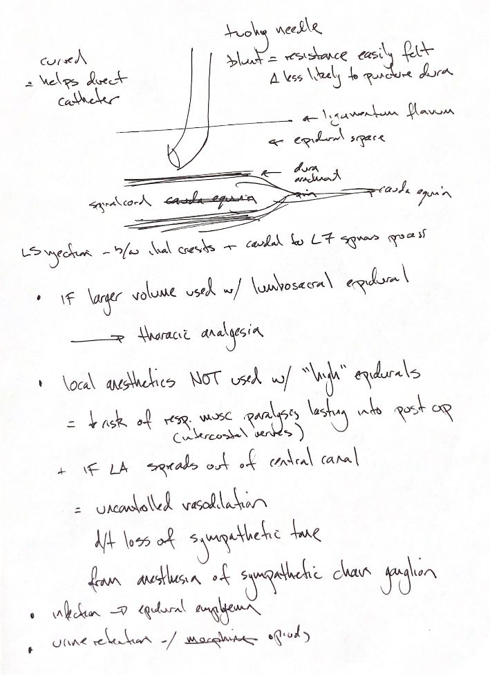
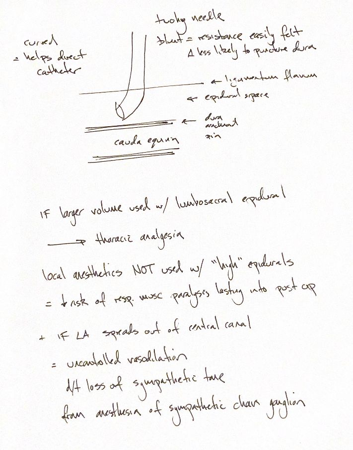
The curved tip of a Tuohy needle helps guide an epidural catheter. It is relatively blunt, but that does not prevent dural puncture. The needle tip belongs in the epidural space, outside the dura. The drawing is a simple layer map, not a guide to needle depth. Cord termination varies with species and age.
My notes place lumbosacral access between the iliac crests, just behind the L7 spinous process. A larger epidural volume can spread farther cranially. Volume alone does not tell us which level will be blocked. The drug, concentration, anatomy and patient position also matter.
The warning about “high” epidurals needs context. The book cautions against long-acting local anaesthetic when wide cranial spread is expected. Intercostal motor block can impair breathing and may last after surgery. This is not a blanket ban on thoracic epidural local anaesthesia.
Sympathetic block can reduce vascular tone and cause vasodilation and hypotension. My notes and the book use “central canal”; the relevant space here is the vertebral canal, not the tiny canal inside the spinal cord. The drug does not have to escape the canal for sympathetic block to occur.
Contamination can cause epidural empyema. Asepsis and preservative-free preparations matter. Epidural opioids, including morphine, can also cause urinary retention. These are separate risks.
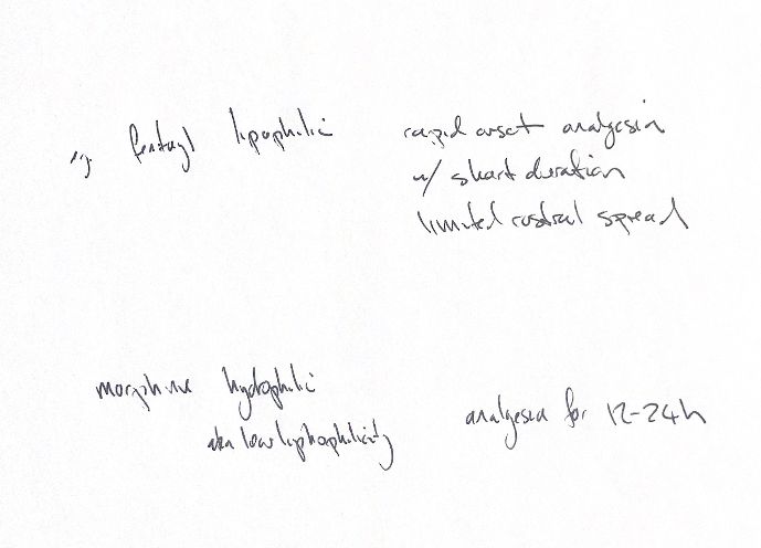
Fentanyl is more lipophilic. Epidural onset is relatively fast, with shorter action and less rostral CSF spread. Morphine is more hydrophilic. It remains in CSF longer and spreads farther rostrally. The 12–24-hour cue fits typical canine pelvic-limb or abdominal use. Duration varies with dose, site and procedure; it is not guaranteed.
References
Johnston & Tobias, 2nd ed., Chapters 21–22; Steagall et al. 2017; Dias et al. 2018.