
Brachial plexus block and nerve origins
Read the draft explanation
The brachial plexus is a network of ventral rami, or branches, of spinal nerves. These branches carry sensory and motor fibres. They are different from the motor-only ventral roots named in my study question.
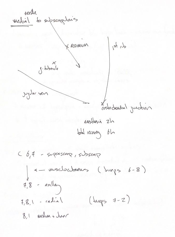
My landmark sketch places the needle medial to subscapularis. It marks the acromion, greater tubercle, jugular vein, first rib and costochondral junction. A published canine nerve-location study describes entry cranial to the acromion and a caudoventral direction medial to subscapularis. The sketch is a conceptual map. It does not name a patient side or give a complete injection technique.
The book describes a brachial plexus block for procedures distal to the elbow. My timing cues say two hours of anaesthesia and six hours to total recovery. The note does not name the drug, dose or measured endpoint. Keep those cues separate from a universal promise about duration.
“SS MARMU” gives the nerve order: suprascapular, subscapular, musculocutaneous, axillary, radial, median and ulnar. The ship is my memory cue. The root lists and muscle groups preserve the supplied chart, including its parenthetical entries.
C6–C7 supplies the main suprascapular and subscapular cues. The axillary cue is C7–C8. The radial list spans C7–T1, with a C7–T2 triceps cue. Median and ulnar share the C8–T1 cue. The supplied chart adds possible contributions, including C5 or T2 in some entries.
Anatomy varies. A study of 18 canine cadavers found 36 plexuses arising from C6–T1, with variation between individual nerves and even between sides of one dog. That sample does not prove that C5 or T2 never contributes.
References
Johnston & Tobias, 2nd ed., Ch22/31/65; Mahler & Adogwa2008; Lambertini2023; University of Minnesota gross anatomy.