
Vasoactive and inotropic agents
Read the draft explanation
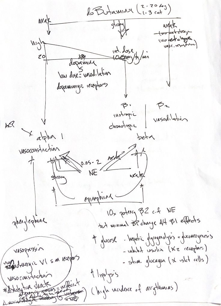
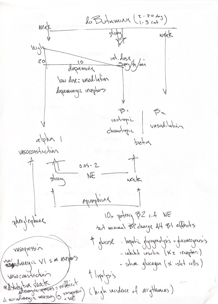
The receptor map links α1 to vasoconstriction, β1 to contractility and heart rate, and β2 to vasodilation. Dobutamine favours β1. The original “no net change” in vascular resistance is a shorthand cue; the response varies with dose and setting.
The dobutamine figures are textbook IV infusion examples in µg/kg/min: 2–20 in dogs and 1–3 in cats. Dopamine’s low, intermediate and high dose groups overlap. Its 5, 10 and 20 cues are not fixed receptor switches. Norepinephrine’s original 0.05–2 range differs from the book’s 0.1–2 and 0.05–0.3 IV infusion tables. These are not one interchangeable regimen.
Phenylephrine favours α1. ACP, read as acepromazine, blocks α1. Norepinephrine has stronger α than β effects, while epinephrine acts at both. My “10× β2 potency versus NE” cue has no verified assay or species basis here. It is not a clinical dose conversion. The “minimal BP change” cue is also conditional: cardiac output and vascular resistance both matter, and epinephrine’s vascular effects change with dose.
Epinephrine raises glucose through hepatic glycogenolysis and gluconeogenesis, reduced insulin release and increased glucagon release. The note’s α2 means an insulin-inhibiting receptor; its α-islet cells are glucagon-producing cells. Lipolysis also rises. Arrhythmias are an important risk, but the drawing gives no measured incidence.
Vasopressin acts through nonadrenergic V1 receptors on vascular smooth muscle. It may help in vasodilatory shock that persists despite adequate volume and catecholamine support. The vasopressin-plus-NE cue is an adjunct concept, not a rule for every distributive shock case.
References
Johnston/Tobias, 2nd ed., Table 6.8 and Chapter 22 drug table; Rosati 2007; Pascoe 2006; Aramaki 2002; Silverstein 2007; official epinephrine label (human physiology context).