
Anticholinergics and vagal events
Read the draft explanation
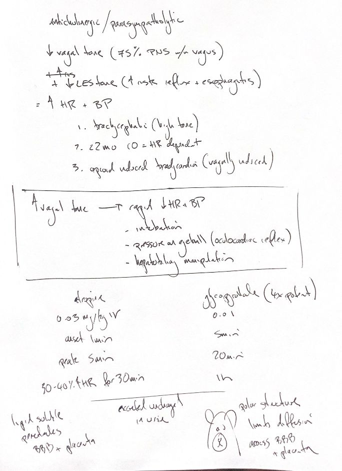
Atropine and glycopyrrolate block muscarinic acetylcholine receptors. They reduce vagal effects on the heart, allowing heart rate to rise. Blood pressure may improve if bradycardia is limiting cardiac output.
They can also reduce lower oesophageal sphincter tone. This may favour reflux and oesophagitis. Pressure and pH measure different effects. A controlled study in 40 dogs found no drug effect on oesophageal, gastric or tracheal pH.
Brachycephalic dogs may have high vagal tone. Very young animals have limited ability to raise stroke volume, so cardiac output depends more on heart rate. My “under two months” cue is a study-note cue, not a validated rule for every puppy or kitten. Opioids can also cause vagal bradycardia.
Intubation, pressure on the eye and hepatobiliary manipulation can provoke a rapid vagal fall in heart rate and blood pressure. Anticipated events call for a patient-specific plan. During an oculocardiac event, stop ocular manipulation while assessing the response. Reflex bradycardia with alpha-2-induced hypertension is a different setting; simply raising heart rate can worsen cardiac work and hypertension.
Atropine acts faster and lasts less time than glycopyrrolate in the reference comparison. Timing varies with route, dose and endpoint. The “four times as potent” summary is not a universal dose-conversion rule. The “75%” vagus cue is a general human-anatomy estimate, not a measured canine proportion or a percentage of vagal tone.
Atropine crosses the blood–brain barrier and placenta. Glycopyrrolate is polar, which limits that passage. Renal elimination matters for both. Atropine is partly metabolised; glycopyrrolate is largely excreted unchanged. Some rabbits have high atropinesterase activity and break down atropine quickly.
References
Johnston & Tobias, 2nd ed., Ch21/124; Grimm et al., Essentials, 2nd ed., pp41–43; Laitinen1978; Roush1990; Ko2001; Harrison2006.