
Nociception and pain processing
Read the draft explanation
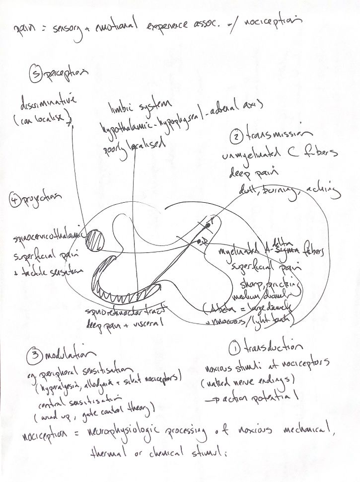
Nociception encodes noxious mechanical, thermal or chemical input. Pain is a sensory and emotional experience. The two are different; a withdrawal reflex alone does not establish conscious pain.
My five study stages are transduction, transmission, modulation, projection and perception. They overlap. Free nociceptor endings turn a noxious stimulus into electrical activity. Nociceptive C fibres are unmyelinated; Aδ fibres are thinly myelinated. The usual dull, burning or aching versus sharp, pricking cues describe qualities, not exclusive tissue routes. Larger Aβ fibres usually carry light touch.
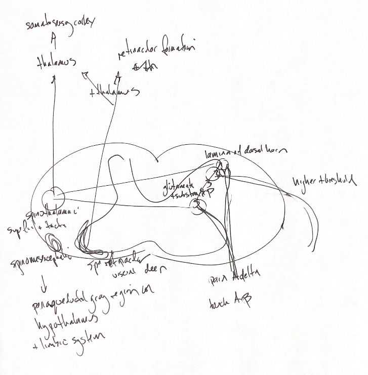
In the dorsal horn, glutamate and substance P help convey nociceptive input. Substance P is not released by every fibre. Laminar connections overlap, so the I/II labels are not a one-fibre, one-layer rule.
Modulation can increase or reduce signalling. Peripheral sensitisation raises nociceptor responsiveness, and some silent nociceptors can be recruited. Hyperalgesia means more pain from a normally painful stimulus; allodynia means pain from normally nonpainful input. Neither proves a purely peripheral mechanism. Central sensitisation, wind-up and gate control are related ideas, but they are not interchangeable.
The first drawing names the spinothalamic tract, with a thalamic relay to somatosensory cortex. Spinomesencephalic input reaches midbrain regions such as the periaqueductal gray, linked to hypothalamic and limbic networks. Spinoreticular input reaches reticular networks.
The second names the spinocervicothalamic route. It first ascends on the same side to the lateral cervical nucleus, before a mainly opposite-side thalamic projection. This route crosses after the cervical relay. These pathways overlap in function. Localisation, emotion and hypothalamic–pituitary–adrenal stress responses involve wider networks.
References
Johnston & Tobias, 2nd ed., Ch26; IASP; cited experimental anatomy.