
Ectopic thyroid and parathyroid tissue
Read the draft explanation
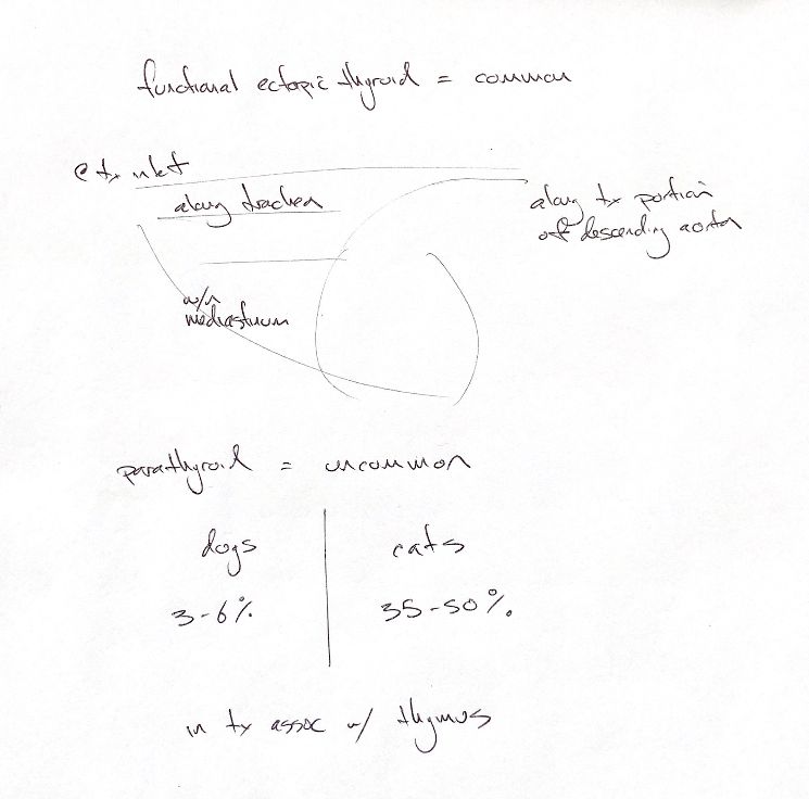
Ectopic means tissue outside its usual location. In dogs and cats, accessory thyroid tissue is common and can be functional. The four sites in this map are along the trachea, at the thoracic inlet, within the mediastinum and along the thoracic part of the descending aorta. Functional tissue can produce thyroid hormone; that alone does not mean it is overactive or neoplastic.
Accessory parathyroid tissue has a different pattern. The textbook reports approximately 3–6% of dogs, with tissue in the thorax associated with the thymus. This is a historical reported estimate, not a modern population prevalence established by these sources.
In cats, older histologic reports describe ectopic parathyroid tissue in 35–50%. Histologic detection means the tissue was identified under the microscope. It does not prove that it can maintain calcium balance after removal of the usual parathyroid glands.
That difference matters. In Flanders and colleagues' experiment, all 14 cats became hypocalcemic after bilateral thyroparathyroidectomy. PTH remained below normal and did not rise significantly during the 12-week study. Tissue presence and immediate functional reserve are separate questions.
References
Johnston & Tobias, 2nd ed., Ch121; Flanders et al. 1991; Marine 1914.