
Thyroid and parathyroid vessels, nerves and lymphatics
Read the draft explanation
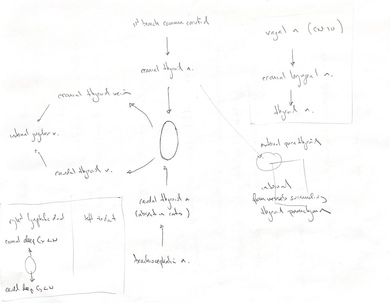
The cranial thyroid artery is the first major branch of the common carotid. It reaches the cranial pole. The caudal thyroid artery usually comes from the brachiocephalic artery and reaches the caudal pole. The arteries connect dorsally, with branching patterns that vary. My cat cue needs “most”: the book describes rare cats with a caudal thyroid artery.
Cranial and caudal thyroid veins leave their respective poles. The source and book show drainage to the internal jugular vein. Individual vessels vary. Some dogs also have an unpaired midline vessel that takes blood from the left gland to the brachiocephalic vein.
The nerve chain is vagus, or cranial nerve X, then cranial laryngeal nerve, then thyroid nerve. The thyroid nerve runs with the cranial thyroid artery. That recall route is not the whole autonomic supply: sympathetic and recurrent-laryngeal contributions have also been described in dogs.
Lymph can leave cranially or caudally through the deep cervical nodes. These nodes may be absent in dogs. Flow may then reach the nearest node, such as the medial retropharyngeal node cranially. The book's terminal cues are the right lymphatic duct and left tracheal duct. Routes vary between animals. Patient left stays on the viewer's right.
External parathyroids receive one or more branches of the cranial thyroid artery. In cats, that branch may start after the artery enters the thyroid capsule. Internal parathyroids receive vessels around the thyroid parenchyma. Venous and lymph drainage, and innervation, share the thyroid routes. The small inset is a supply sketch, not a fixed map of gland positions.
References
Johnston & Tobias, 2nd ed., Ch121; anatomical checks include Stocco 2021 and Mizeres 1955.