
GI and colonic wound healing versus skin
Read the draft explanation
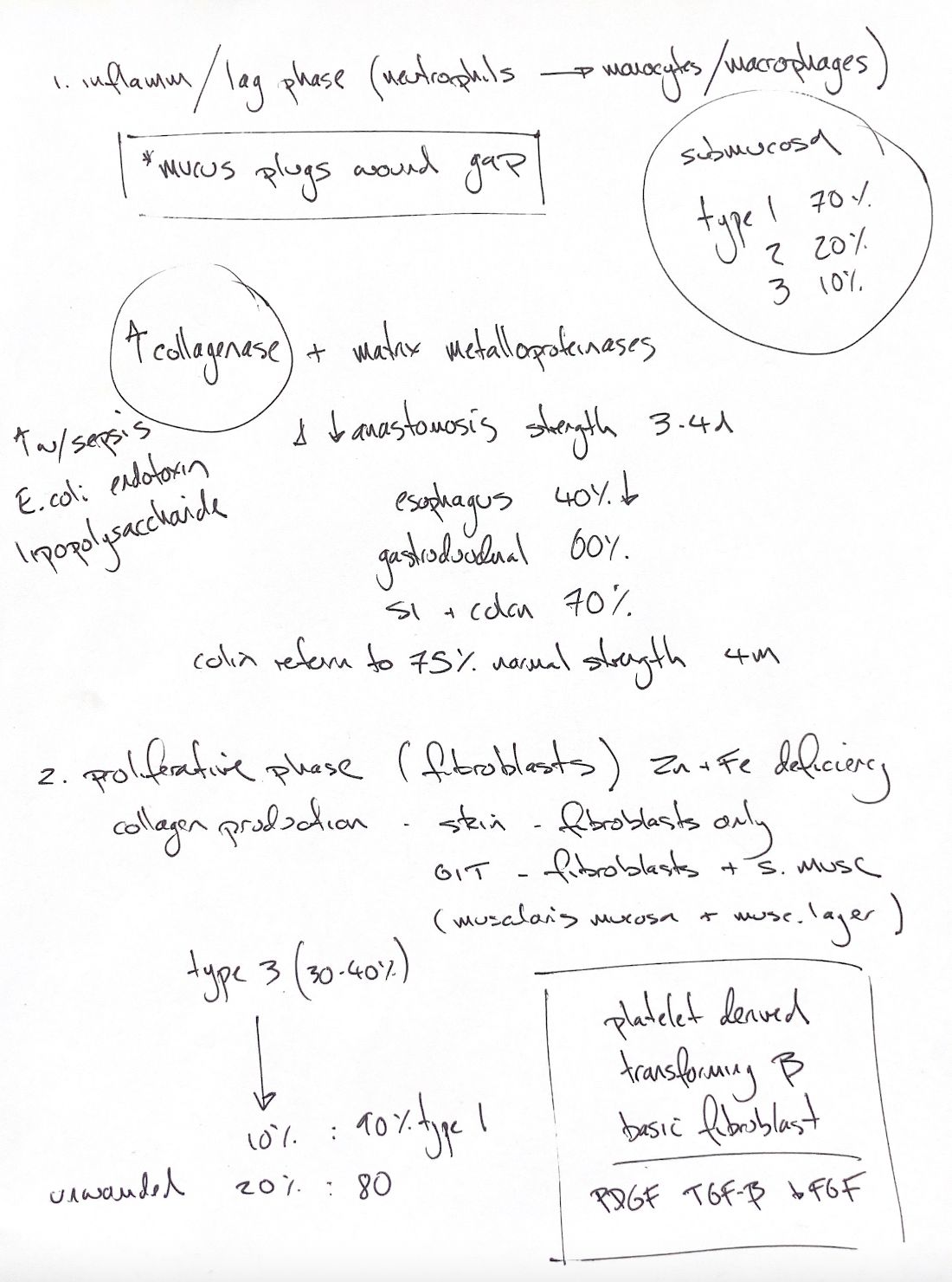
The colon follows the same overlapping healing phases as skin. The early difference is weakness. Neutrophils arrive first; monocytes and macrophages then predominate. Mucus and fibrin help seal the gap, while sutures provide the critical support.
Collagenase and other matrix metalloproteinases break down collagen. Sepsis can increase loss and reduce production. E.coli lipopolysaccharide can stimulate collagenase synthesis in activated macrophages. The first 3–4 days are a vulnerable period.
My rounded early loss cues are 40% oesophageal, 60% gastroduodenal and 70% intestinal. The rat primary study measured suture holding against the immediate postsuture baseline: losses of 37%, 64% and 72% at 48 hours, but 70% for small intestine at 24 hours. These are not leak rates or dog/cat predictions.
The submucosa supplies most GI tensile support. My rounded 70/20/10 circle needs clearer type labels: the book gives collagen I 68%, III 20% and V 12%.
Skin wound collagen comes mainly from fibroblasts. In the gut, fibroblasts and smooth muscle cells contribute, including muscle in the muscularis mucosae and propria. PDGF, TGF-β and basic FGF support proliferation. Experimental zinc or iron deficiency reduces fibroblast activity and collagen production.
Type III is prominent early, then its proportion falls during remodelling. The note’s 20% III/80% I unwounded cue and 10% III/90% I later cue match the book’s dermal comparison. They are not a complete I/III/V composition of GI submucosa.
The note and book text give about 75% normal colonic strength at 4 months. Other book descriptions use earlier near-normal bursting strength or a lower breaking-strength curve. Different tests and baselines cannot be merged into one recovery timetable.
References
Johnston & Tobias, 2nd ed., Ch9/93; Högström & Haglund 1985 (PMID3911698).