
Gastric glands and secretory cells
Read the draft explanation
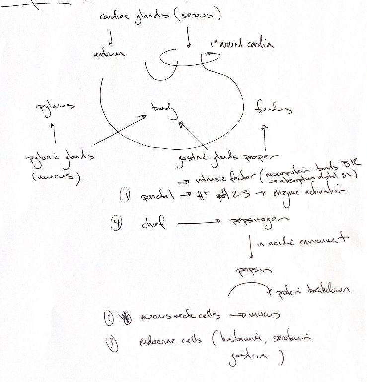
My region map follows the supplied book: cardiac glands around the cardia and also in the antrum; pyloric glands in the pylorus and body; gastric glands proper in the fundus and body. These are source descriptions, not sharp universal boundaries.
The source and book call cardiac secretion “serous”. This needs qualification. Dog and cat histology shows mixed secretory features, while teaching guides often describe mucus. The source label is not a rule that these glands are purely serous. Pyloric glands mainly produce mucus.
My four cell cues keep their original numbers: 1 parietal, 4 chief, 2 mucous neck and 3 endocrine.
Parietal cells pump H+ into the gastric lumen. The source’s pH 2–3 is an acidic memory cue, not a fixed value in every animal or feeding state. Acid helps activate digestive enzymes.
The intrinsic-factor cue needs a species limit. Dogs make most intrinsic factor in the pancreas, with a smaller gastric contribution that includes parietal cells. Cats rely on pancreatic intrinsic factor. This glycoprotein, also called a mucoprotein in the note, binds vitamin B12 and supports uptake in the distal small intestine, mainly the ileum.
Chief cells secrete inactive pepsinogen. In the acidic lumen it becomes pepsin, which breaks down proteins. The cell secretes the precursor; activation occurs outside it. Mucous neck cells secrete protective mucus.
The endocrine list contains histamine, serotonin and gastrin. These do not all come from one cell or share one route. Gastrin-producing G cells are mainly in the pyloric antrum in dogs, so the book table’s “body” label is not a location rule for the whole list. Histamine can act locally as a paracrine signal. Serotonin is associated with enterochromaffin cells. These signals do not all enter the gastric lumen.
References
Johnston & Tobias, Veterinary Surgery: Small Animal, 2nd ed.; primary dog/cat studies and university histology guides.