
Oesophageal sphincters and blood supply
Read the draft explanation
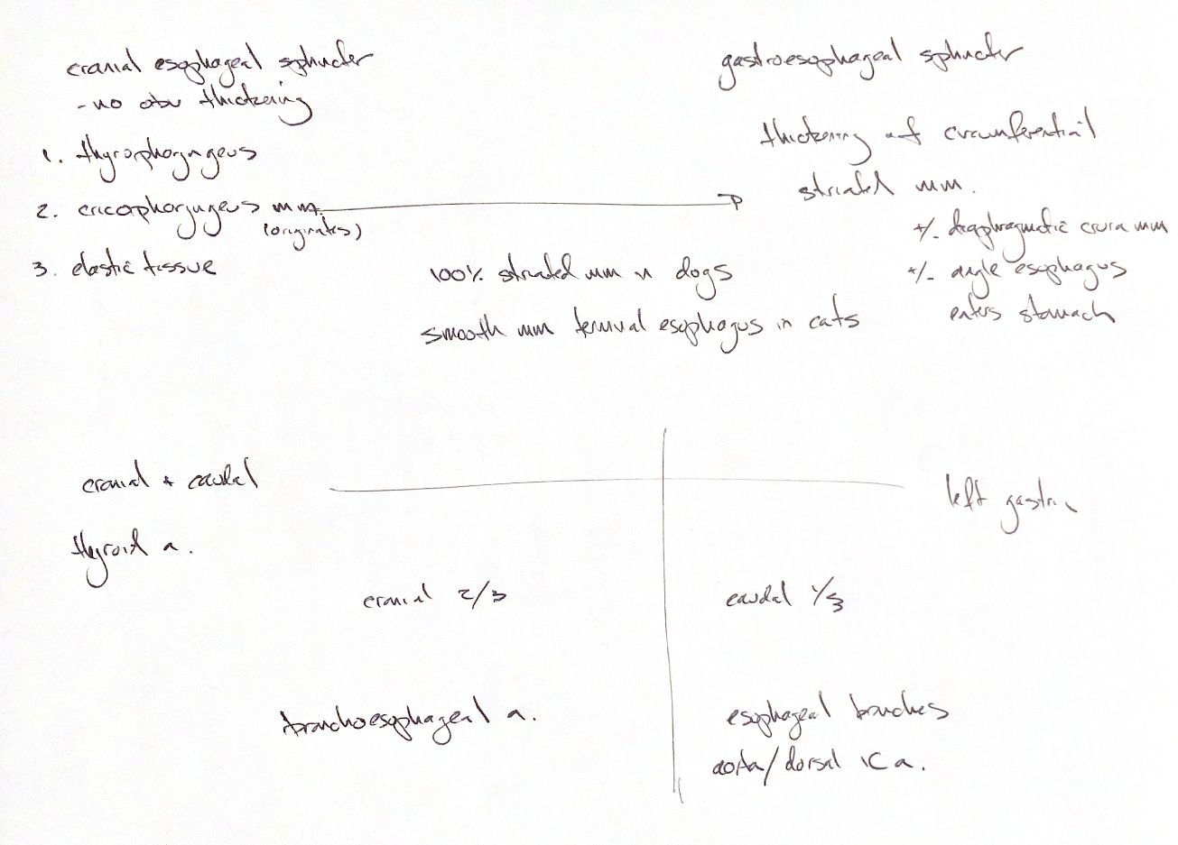
The cranial oesophageal sphincter has no obvious wall thickening. Thyropharyngeus, cricopharyngeus and elastic tissue provide its function. The oesophageal muscularis starts from cricopharyngeus and the cricoesophageal tendon.
In dogs, this muscularis is striated along its full length. Cats have smooth muscle in the terminal oesophagus. In dogs, circumferential striated-muscle thickening at the gastroesophageal junction may mark the caudal sphincter. The diaphragmatic crura and entry angle are proposed contributors, as the ± cues in my note suggest. These functional sphincters have boundaries that are hard to define anatomically.
Follow the arterial map from cranial to caudal. My note lists cranial and caudal thyroid branches for the cervical region. The caudal thyroid artery is absent in most cats. The bronchoesophageal artery is the main supply to the cranial two-thirds of the THORACIC oesophagus. Oesophageal branches of the aorta or dorsal intercostal arteries supply the remaining third. A left gastric branch supplies the terminal portion. This is a regional recall map; individual arterial origins vary.
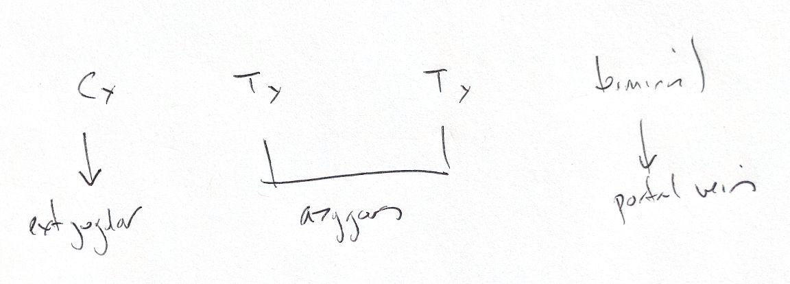
Venous blood drains from the cervical part to the external jugular veins, from the thoracic part mostly to the azygos vein, and from the terminal part via the vein accompanying the left gastric artery into the portal system. A rich submucosal vascular plexus links the segmental vessels.
References
Johnston & Tobias, 2nd ed., Ch90/121; Resoagli et al.2006.