
Thromboembolism and thromboelastography
Read the draft explanation
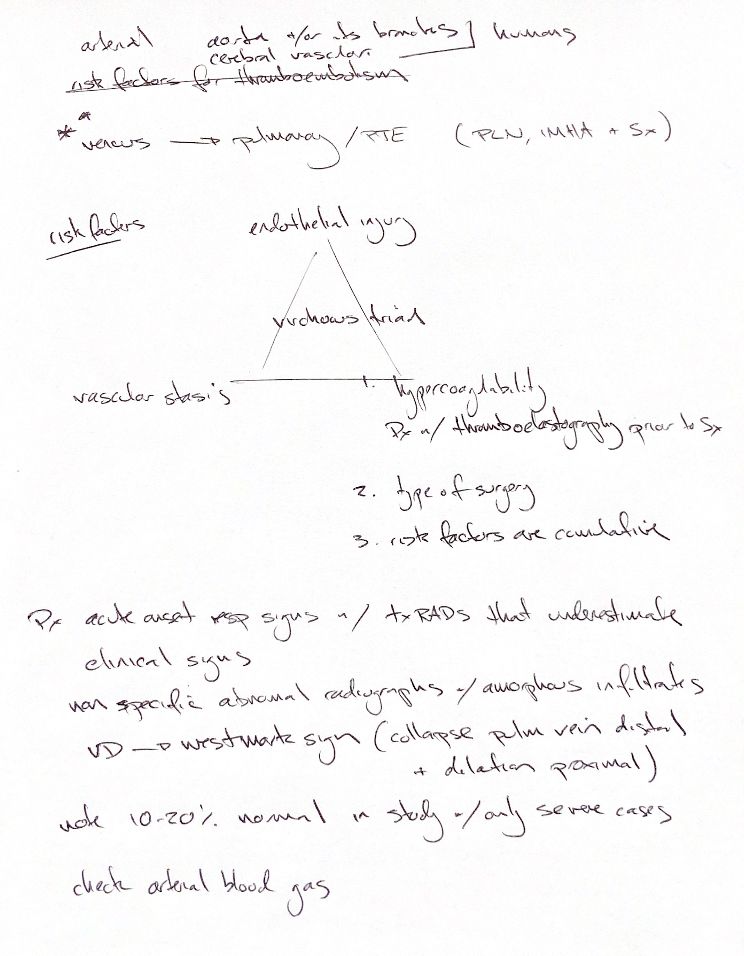
Virchow's triad links endothelial injury, vascular stasis and hypercoagulability. Risks add together, including the type of surgery. In small animals, arterial thrombi often involve the aorta, its branches or cerebral vessels. Venous disease often affects the lungs. Protein-losing nephropathy, IMHA and surgery are useful PTE risk cues; the arterial and venous associations overlap.
Suspect pulmonary thromboembolism (PTE) when acute respiratory distress exceeds the changes on chest radiographs. Infiltrates can be amorphous and nonspecific. The Westermark sign is distal oligemia, best seen on VD or DV views. An attenuated lobar artery or vein and a large main pulmonary artery are possible changes, not a required diagnostic pair. Blood gases assess gas exchange but do not confirm PTE.
The note gives 10–20% normal radiographs. The supplied book gives 9–27% in dogs from necropsy series, which select more severe cases. In one study, 6 of 22 radiographed dogs had normal films. Normal films do not exclude PTE.
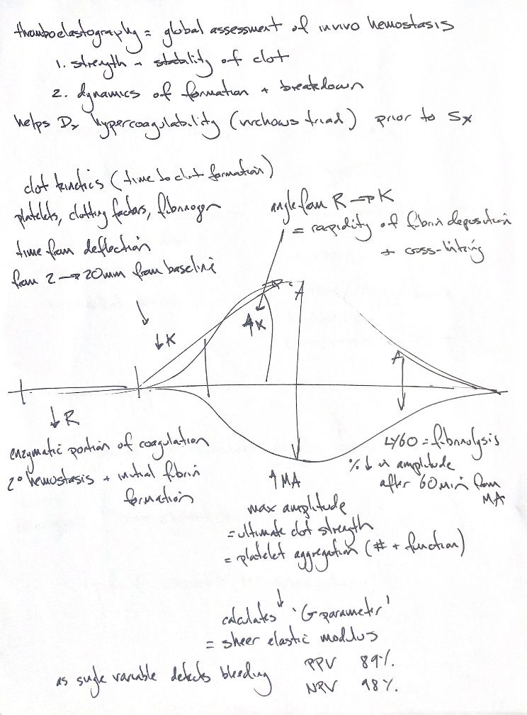
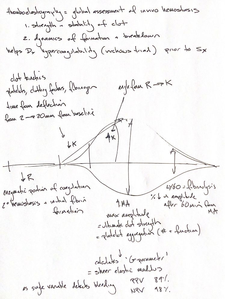
TEG tests whole blood outside the body. R marks initial fibrin formation at 2 mm trace width. K runs from R until 20 mm. The α-angle reflects fibrin build-up and cross-linking. MA shows clot strength, mainly from platelets and fibrinogen. G is derived from MA. LY60 describes the percentage fall in amplitude 60 minutes after MA.
A hypercoagulable pattern has higher G and may have shorter R/K or higher MA/α. Compare with the same species and assay protocol. The trace does not prove thrombosis or guarantee a surgical outcome. In a selected canine TF-activated TEG study, G below 3.2 kdyn/cm² identified concurrent clinical bleeding with PPV 89% and NPV 98%. These study values are not universal cutoffs.
References
Johnston & Tobias, 2nd ed., Ch. 7; Johnson 1999; Wiinberg 2009; Thawley 2016.