
Stem-cell origins and potency
Read the draft explanation
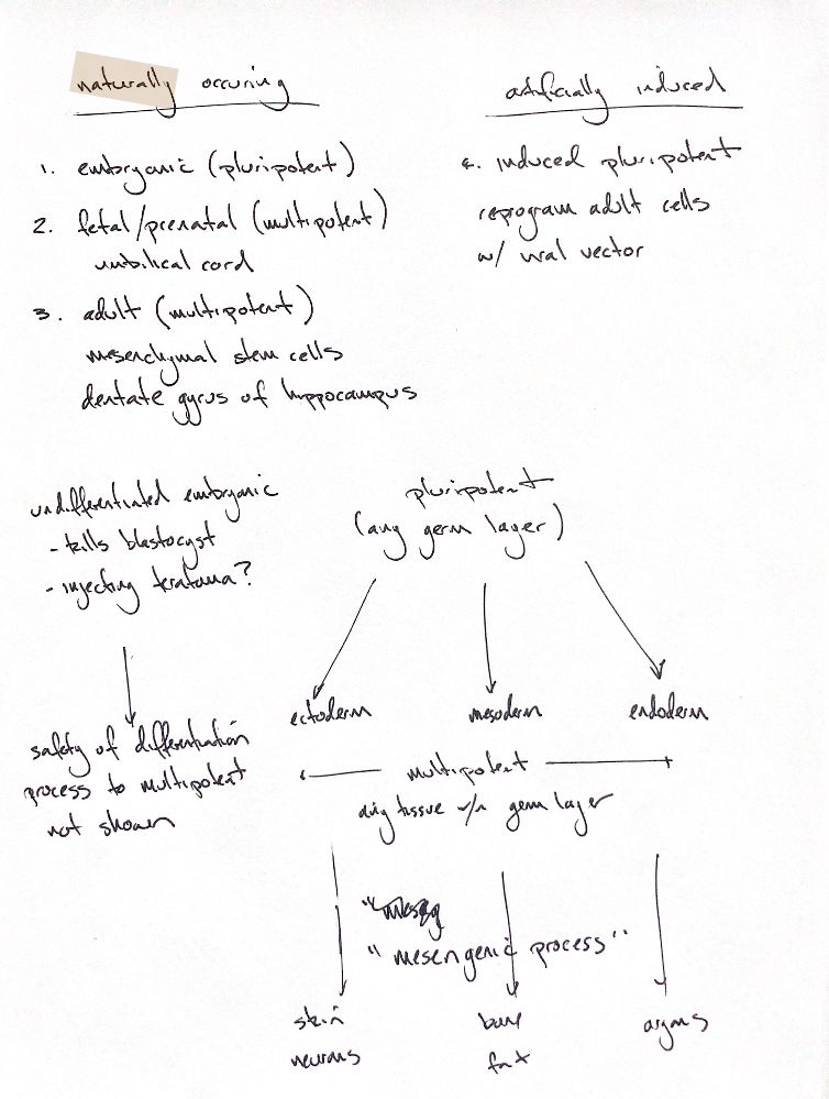
The two broad groups here describe origin: naturally occurring stem cells and artificially induced cells. Potency describes which cell types they can form.
Naturally occurring embryonic stem cells are pluripotent. They can form cells from ectoderm, mesoderm and endoderm, but do not have the full extraembryonic potential of a totipotent zygote. The original “fetal/prenatal” group includes umbilical-cord cells, usually classed as perinatal. These cells are generally multipotent. Adult stem cells are also usually multipotent. Mesenchymal stem cells are one example. Neural stem cells are another; the dentate gyrus of the hippocampus is a neural stem-cell niche.
Induced pluripotent stem cells, or iPSCs, start as adult somatic cells. Reprogramming resets their gene-expression programme toward a pluripotent state. Viral vectors are one way to do this, but nonviral methods also exist.
The lineage tree shows increasing restriction. Multipotent cells produce several specialised cell types within a lineage. Skin cells and neurons illustrate ectodermal derivatives. Bone and fat illustrate mesodermal derivatives. The endodermal “organs” cue refers to structures such as gut and liver epithelium; whole organs contain cells from several lineages. These examples come from different tissue-specific stem-cell pools. The mesengenic process specifically describes MSC lineage commitment and differentiation, rather than every branch of the tree.
Conventional embryonic stem-cell derivation removes the blastocyst's inner cell mass and destroys the embryo. Undifferentiated pluripotent cells can form teratomas after transplantation into immunocompromised animals. These tumours contain disordered mixtures of differentiated tissues. Directing cells toward the desired lineage aims to reduce that risk. Differentiation alone does not establish clinical safety for a particular product and use.
References
Johnston/Tobias, Veterinary Surgery: Small Animal, 2nd ed., Ch. 3; NIH Stem Cell Basics; Takahashi & Yamanaka, Cell 2006;126:663–676; Reubinoff et al., Nat Biotechnol 2000;18:399–404.