
Pancreatic cells, products, protection and canine ducts
Read the draft explanation
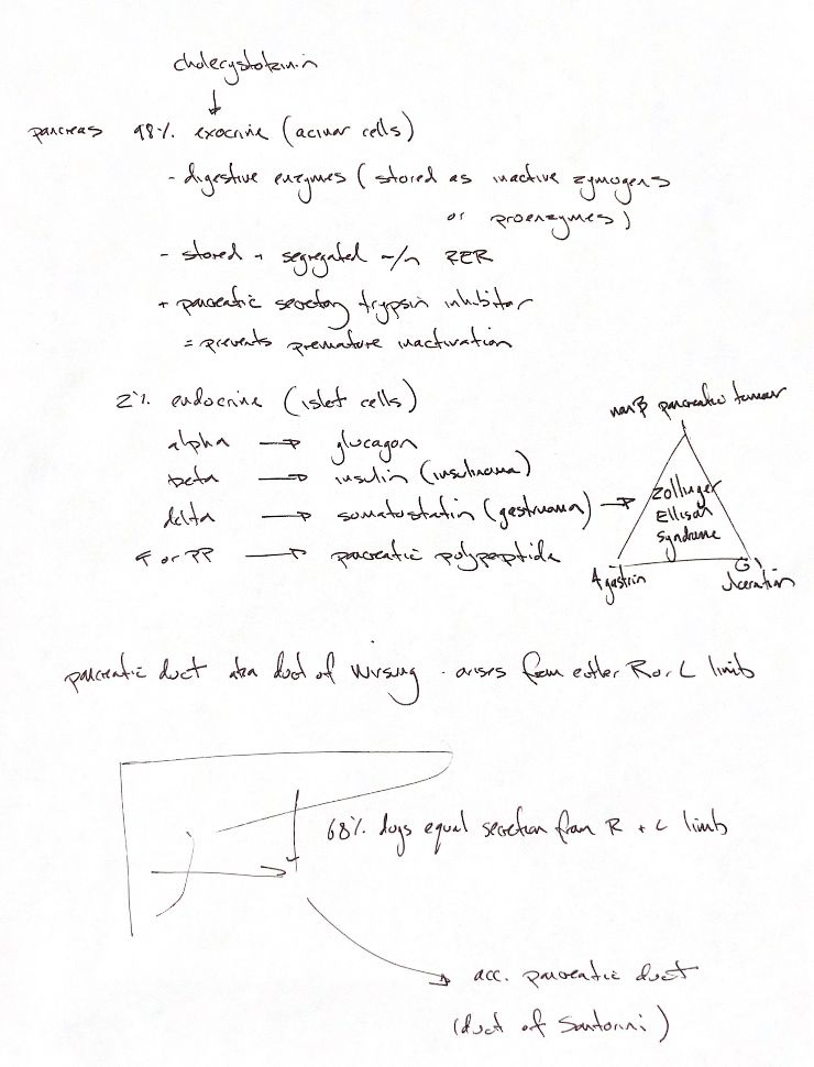
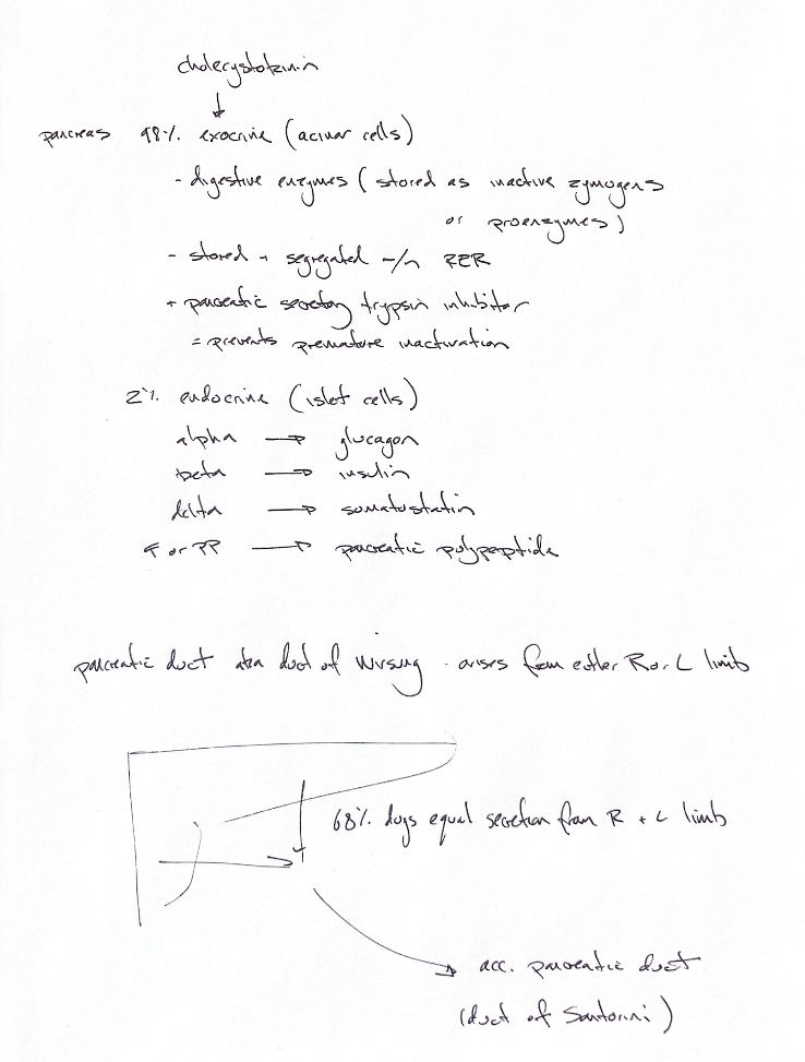
Most pancreatic mass is exocrine. The 98% cue includes acinar tissue, ducts, vessels and nerves. Roughly 2% is endocrine. These are mass estimates, not cell counts. Acinar cells send digestive enzymes through ducts. Cholecystokinin, or CCK, stimulates enzyme release.
Several safeguards limit autodigestion. Proteases are made as inactive zymogens and kept in separate granules. Amylase and lipase are secreted active, so “all enzymes are inactive” would be wrong. Pancreatic secretory trypsin inhibitor inhibits trypsin and limits premature activation. The handwriting appears to say “premature inactivation”; the checked mechanism prevents activation. The original and book place stored granules within rough endoplasmic reticulum. That needs correction: RER makes proteins, the Golgi processes them, and apical granules store them before release.
Islets release hormones into blood. Alpha cells make glucagon. Beta cells make insulin; neoplastic beta cells form insulinomas. Delta cells make somatostatin. F, or PP, cells make pancreatic polypeptide.
The original links delta cells to gastrinoma. Keep this as a proposed conversion model, since exact tumour-cell lineage remains uncertain. Normal gastrin comes from gastrointestinal G cells. The classic Zollinger–Ellison triangle joins a non-beta pancreatic tumour, high gastrin and GI ulceration. Excess gastrin raises gastric acid. This is a classic pattern, not a claim that every gastrinoma causes ulcers.
In the book’s reported 68% canine pattern, ducts from both pancreatic limbs form a Y. Its tail is the larger accessory duct, or Santorini, which reaches the minor duodenal papilla and carries most secretion. A smaller pancreatic duct, or Wirsung, may arise from either limb. It reaches the major papilla beside the bile duct. Individual anatomy varies. The 68% counts duct patterns; it does not prove equal right/left secretion. Its original primary table was not obtained.
References
Chris Wood’s two drawings; Johnston & Tobias, Veterinary Surgery: Small Animal, 2nd ed., ch97; UMN Histology Guide, acinar-cell TEM; Eddeland & Ohlsson 1978, PMID81511; Gal et al. 2011; Struthers et al. 2018, PMID29860931 (lineage discussion).