
Canine portal drainage and the variable colic junction
Read the draft explanation
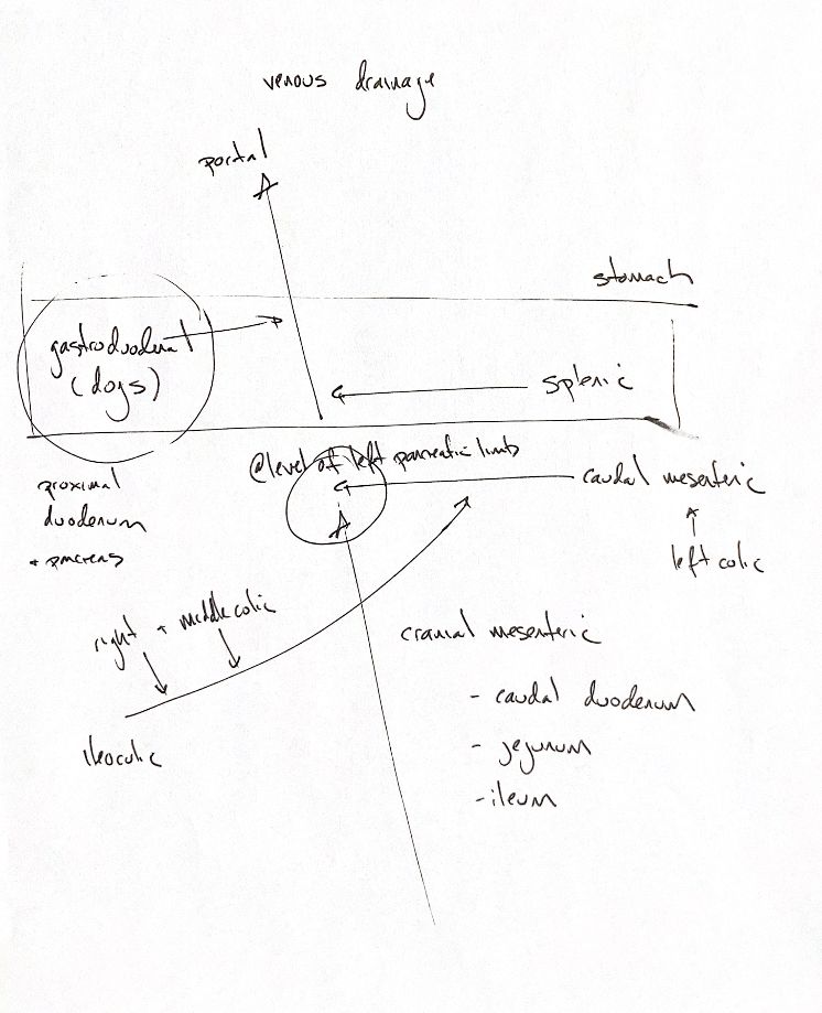
Follow the arrows toward the liver. This original sketch maps portal drainage from selected abdominal viscera. It is not a map of every abdominal vein.
The cranial mesenteric vein collects blood from the caudal duodenum, jejunum and ileum. It joins the caudal mesenteric route to form the portal vein. The source marks this junction at the level of the left pancreatic limb. Keep that as a memory landmark, not a measured distance.
The left colic vein drains into the caudal mesenteric vein. In the original map, right and middle colic veins join the ileocolic vein, which then joins the caudal mesenteric route. The oblique line crosses the cranial mesenteric line on the page, but that crossing is not another junction.
This colic connection needs a qualifier. Primary dissection in eight Kangal dogs included ileocolic drainage into the caudal mesenteric vein. Other descriptions group these vessels with cranial mesenteric drainage. Exact junctions vary, so the original route is a useful recall map rather than a universal branch template. The textbook’s colonic prose also describes middle colic drainage in more than one place.
More cranially, the splenic vein joins the portal vein. The stomach drains through the splenic route on the left and the gastroduodenal route on the right. In the dog, the gastroduodenal vein also drains the proximal duodenum and parts of the pancreas before joining the portal vein. A dog-specific label here does not establish that this vein is absent in cats.
References
Johnston/Tobias 2e, small-intestinal, colonic and hepatic vascular chapters; Kalt & Stump1993; Dursun et al.1994. The small primary dissection series supports variation; it does not give a general population rate.